The arterioles, as the smallest branches of the arterial system, play a crucial role in regulating blood flow from arteries to capillaries, adapting to the body’s immediate metabolic needs. This image highlights the tunica intima, tunica media, tunica adventitia, and smooth muscle cells, showcasing the structural features that allow these tiny vessels to control peripheral resistance and capillary perfusion.
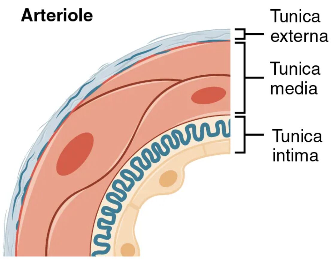
Tunica intima Tunica intima is the innermost layer, consisting of a thin endothelial cell lining that facilitates blood flow into capillaries. This layer includes a minimal subendothelial layer and lacks a prominent elastic lamina, reflecting the arteriole’s role in fine-tuned flow regulation rather than high-pressure resistance.
Tunica media Tunica media forms a thin middle layer, composed of one or two layers of circular smooth muscle cells with very few elastic fibers. This structure enables the arteriole to constrict or dilate in response to local metabolic signals, controlling the amount of blood entering capillary beds.
Tunica adventitia Tunica adventitia is the outermost layer, made up of a sparse network of connective tissue that anchors the arteriole to surrounding tissues. This thin layer provides minimal structural support and lacks significant vasa vasorum, as the vessel’s small size reduces the need for extensive nourishment.
Smooth muscle cells Smooth muscle cells dominate the tunica media, appearing as a thin ring of contractile cells under magnification, and are responsible for adjusting the arteriole’s diameter. These cells respond to local factors like oxygen levels and pH, as well as neural and hormonal inputs, to regulate blood flow and pressure.
The Role of Arteriolar Layers in Circulation
This overview highlights how each layer supports the arteriole’s regulatory function. The design ensures precise control of blood distribution to tissues.
- Tunica Intima Function: The endothelial cells release nitric oxide to promote vasodilation, enhancing capillary flow. The thin structure minimizes resistance, aiding nutrient exchange.
- Tunica Media Role: The smooth muscle cells contract to increase resistance or relax to increase flow, responding to metabolic demands. This layer’s thinness allows rapid adjustments.
- Tunica Adventitia Support: The connective tissue stabilizes the arteriole, preventing collapse under pressure changes. Its simplicity reflects the vessel’s small size and short length.
- Smooth Muscle Cell Activity: These cells adjust lumen size based on local tissue needs, such as during exercise. Their responsiveness maintains optimal capillary perfusion.
Anatomical Details of Arterioles
The image provides a detailed view of the arteriole’s layered structure, emphasizing its simplicity. The thin tunica media stands out in this design.
This close-up perspective aids in understanding the vessel’s regulatory role. It serves as a foundation for studying microcirculation and vascular control.
- Tunica Intima Composition: The endothelium is supported by a minimal subendothelial layer, with no significant elastic lamina. This thinness optimizes flow into capillaries.
- Tunica Media Thickness: The layer contains one or two layers of smooth muscle, with scant elastic fibers. This structure is tailored for dynamic diameter changes.
- Tunica Adventitia Variation: The outer layer is a thin collagen network, lacking prominent vasa vasorum. Its simplicity supports the arteriole’s small diameter.
- Smooth Muscle Cell Structure: These cells form a sparse, circular arrangement around the lumen. Their density decreases as the arteriole nears capillary junctions.
Physiological Functions of Arteriolar Layers
The physiological roles of these layers are designed to manage blood flow at the microcirculatory level. Their design ensures effective tissue perfusion.
Each layer contributes uniquely to arteriole performance. This functionality supports the body’s cardiovascular balance.
- Blood Flow Regulation: The tunica media’s smooth muscle adjusts resistance, contributing to about 50% of total peripheral resistance. This control directs blood to active tissues.
- Pressure Control: The thin walls allow pressure to drop from arterial levels to capillary levels, around 35 mmHg. This gradient facilitates exchange in capillary beds.
- Oxygen Delivery: Arterioles carry oxygenated blood, with the tunica intima’s smooth surface ensuring efficient flow. This supports oxygen delivery to metabolizing tissues.
- Metabolic Response: Smooth muscle cells dilate in response to low oxygen or high carbon dioxide, increasing flow. This autoregulation matches blood supply to demand.
Comparative Anatomy with Other Arterial Types
The arteriole contrasts with elastic and muscular arteries, reflecting its unique microcirculatory role. This image emphasizes these structural differences.
The visual representation highlights the arteriole’s distinct features. This understanding is key to grasping vascular diversity.
- Arteriole vs. Elastic Arteries: Arterioles have a thin tunica media with minimal elastic fibers, unlike the elastic lamellae-rich media of elastic arteries. This reflects their role in resistance versus pressure maintenance.
- Arteriole vs. Muscular Arteries: Arterioles feature a thinner media with fewer muscle layers, contrasting with the thick, muscular media of muscular arteries. This shift supports finer flow control.
- Transition to Capillaries: The tunica media thins into precapillary sphincters, reducing to a single endothelial layer. This transition maximizes exchange surfaces.
- Histological Features: Staining reveals sparse smooth muscle in the tunica media, contrasting with the elastic or muscular focus in larger arteries. This aids in microscopic identification.
Clinical and Research Perspectives
Insights from arteriolar anatomy inform medical practice and research. The layered structure is a focus for studying microcirculatory health and disease.
Advances in imaging and histology enhance these studies, offering diagnostic tools. These efforts support innovative treatment strategies.
- Hypertension Impact: Narrowed arterioles due to smooth muscle hypertrophy increase resistance. Microscopic analysis guides antihypertensive therapy.
- Diabetic Microangiopathy: Thickened tunica media in arterioles reduces flow, visible under microscopy. This finding directs glucose management.
- Peripheral Vascular Disease: Reduced arteriole dilation affects limb perfusion, detectable via imaging. Early intervention improves outcomes.
- Therapeutic Innovations: Targeting smooth muscle with vasodilators treats hypertension. Stem cell research explores regenerating arteriole layers.
In conclusion, this image of an arteriole provides a detailed look at the tunica intima, tunica media, tunica adventitia, and smooth muscle cells, revealing their critical roles in regulating blood flow to capillaries. These anatomical insights not only deepen our understanding of microcirculation but also support advancements in diagnosing and treating vascular conditions.

