Capillaries are the smallest blood vessels in the body, playing a crucial role in the exchange of nutrients, gases, and waste products between blood and tissues. This diagram illustrates the three major types—continuous, fenestrated, and sinusoid—highlighting their unique structural features that determine permeability and function in various organs.
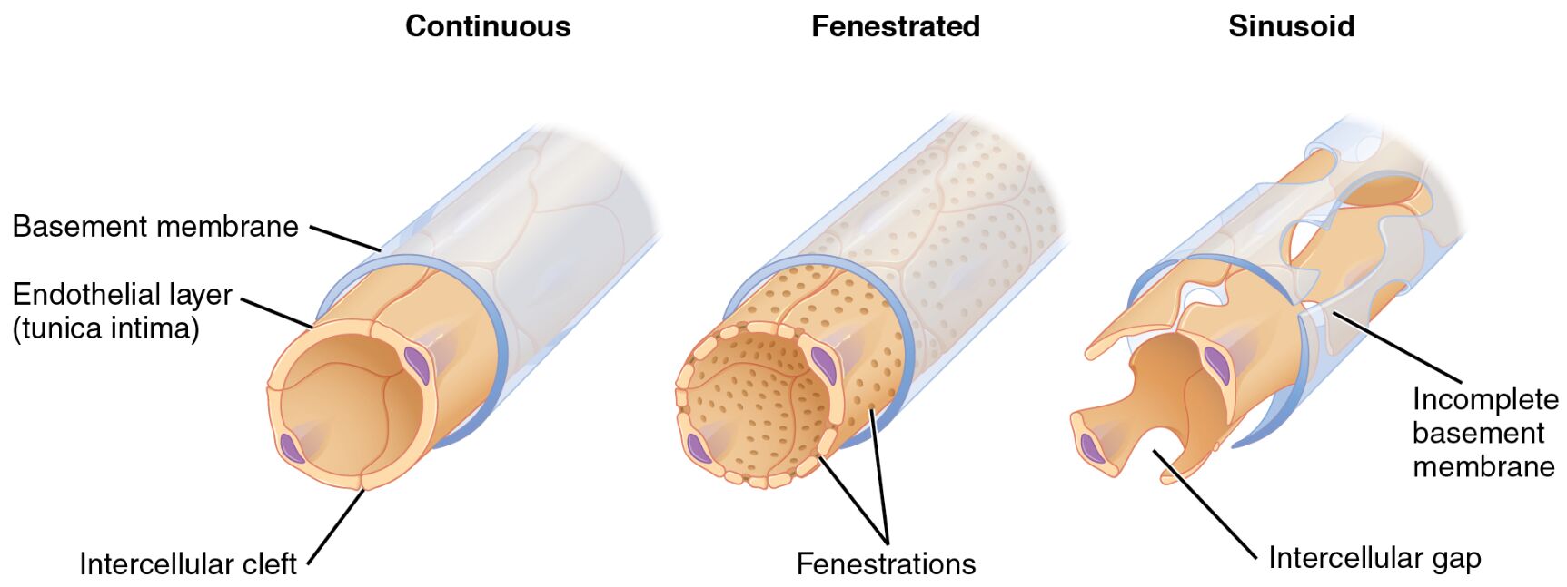
Basement membrane The basement membrane is a thin layer of extracellular matrix that provides structural support to the endothelial cells in capillaries. It acts as a selective barrier, regulating the passage of molecules and contributing to the overall integrity of the vessel wall.
Endothelial layer (tunica intima) The endothelial layer, also known as the tunica intima, consists of a single layer of flattened endothelial cells that line the interior of blood vessels. This layer is essential for maintaining blood flow, preventing clotting, and facilitating the exchange processes in capillaries.
Intercellular cleft The intercellular cleft refers to the narrow space between adjacent endothelial cells in continuous capillaries. It allows for the diffusion of small molecules like water and ions while restricting larger substances, thus controlling permeability.
Fenestrations Fenestrations are small pores or openings in the endothelial cells of fenestrated capillaries. These pores enhance the vessel’s permeability, enabling rapid exchange of fluids and solutes in organs requiring high filtration rates.
Incomplete basement membrane The incomplete basement membrane in sinusoid capillaries features gaps or discontinuities, allowing for greater permeability. This structure supports the passage of larger molecules and cells, which is vital in organs involved in blood filtration and storage.
Intercellular gap The intercellular gap in sinusoid capillaries is a wider space between endothelial cells compared to other types. It facilitates the easy movement of blood cells and large proteins, making these capillaries highly permeable for specialized functions.
The Role of Capillaries in the Circulatory System
Capillaries serve as the vital link between arteries and veins, ensuring efficient microcirculation throughout the body. Their diverse structures adapt to the specific needs of different tissues, from tight barriers in sensitive areas to highly permeable walls in absorptive regions.
- Capillaries form extensive networks, with their total surface area in the human body estimated at around 600 to 700 square meters, optimizing exchange processes.
- They receive oxygenated blood from arterioles and return deoxygenated blood to venules, maintaining homeostasis.
- Structural variations among capillary types influence how substances like oxygen, carbon dioxide, and hormones such as T3 and T4 from the thyroid gland are transported to target cells.
- In response to local demands, capillaries can dilate or constrict through mechanisms involving endothelial-derived factors like nitric oxide.
Structural Differences Among Capillary Types
Each type of capillary exhibits distinct endothelial arrangements and basement membrane characteristics that suit their physiological roles. These differences are critical for understanding organ-specific functions and potential pathologies.
- Continuous capillaries feature a complete endothelial lining with tight junctions, minimizing leakage.
- Fenestrated capillaries include pores that allow selective permeability, ideal for filtration.
- Sinusoid capillaries have discontinuous walls, permitting the passage of cells like red blood cells.
- The basement membrane varies from continuous and intact to fragmented, affecting molecular sieving.
Continuous Capillaries: The Standard Model
Continuous capillaries are the most common type found in muscles, skin, and the central nervous system. They provide a controlled environment for diffusion while protecting delicate tissues from excessive fluid loss.
- Their endothelial cells are joined by tight junctions, with only small intercellular clefts for passage.
- The complete basement membrane acts as an additional filter, preventing large proteins from escaping.
- In the brain, these form the blood-brain barrier, selectively allowing nutrients while blocking toxins.
- They support slow, steady exchange suitable for tissues with consistent metabolic needs.
Fenestrated Capillaries: Enhanced Permeability
Fenestrated capillaries are specialized for rapid fluid and solute exchange, commonly located in endocrine glands and kidneys. Their pores, or fenestrations, measure about 60-80 nanometers in diameter, facilitating filtration without compromising structural integrity.
- Found in the glomeruli of kidneys, they enable the formation of urine by filtering blood plasma.
- In intestinal villi, they aid in nutrient absorption from digested food.
- Endocrine organs like the pancreas utilize them for hormone secretion into the bloodstream.
- The basement membrane remains continuous, providing support despite the pores.
Sinusoid Capillaries: Maximum Exchange
Sinusoid capillaries, also known as discontinuous capillaries, are designed for maximal permeability and are present in the liver, spleen, and bone marrow. Their irregular, widened lumens and gaps allow for the direct interaction between blood and surrounding cells.
- In the liver, they permit hepatocytes to access plasma proteins and remove toxins efficiently.
- The spleen uses them for filtering old red blood cells and immune surveillance.
- Bone marrow sinusoids facilitate the release of new blood cells into circulation.
- Incomplete basement membranes and large intercellular gaps distinguish them from other types.
Physiological Importance of Capillary Variations
The adaptability of capillary structures ensures that each organ receives precisely what it needs for optimal function. Disruptions in these structures can lead to conditions like edema or impaired nutrient delivery, underscoring their importance in health.
- Continuous capillaries maintain barrier functions in protective environments like the lungs.
- Fenestrated types support high-volume exchanges in absorptive and secretory tissues.
- Sinusoids enable cellular trafficking, crucial for hematopoiesis and detoxification.
- Overall, these variations contribute to the body’s ability to regulate blood pressure and fluid balance.
Clinical Relevance and Applications
Understanding capillary types aids in diagnosing and treating vascular-related disorders. For instance, alterations in permeability can indicate inflammation or disease processes.
- In diabetes, damage to endothelial layers can lead to microvascular complications.
- Therapies targeting fenestrations aim to improve drug delivery in cancer treatment.
- Sinusoid dysfunction in liver cirrhosis affects blood flow and detoxification.
- Research into capillary regeneration holds promise for wound healing and tissue engineering.
In summary, the diagram of continuous, fenestrated, and sinusoid capillaries provides a clear visual representation of how structural adaptations enable specialized functions in the vascular system. By appreciating these differences, one gains deeper insight into the intricate balance of exchange and protection that sustains life at the cellular level.

