The regulation of water balance in the human body is a finely tuned process, with the kidneys playing a central role. This diagram illustrates the critical function of aquaporins in the collecting tubules of the kidney, detailing how these specialized water channels facilitate the reabsorption of water from the filtrate back into the bloodstream. This mechanism is profoundly influenced by Antidiuretic Hormone (ADH) and is essential for maintaining proper hydration and blood volume.
Understanding Aquaporin Function
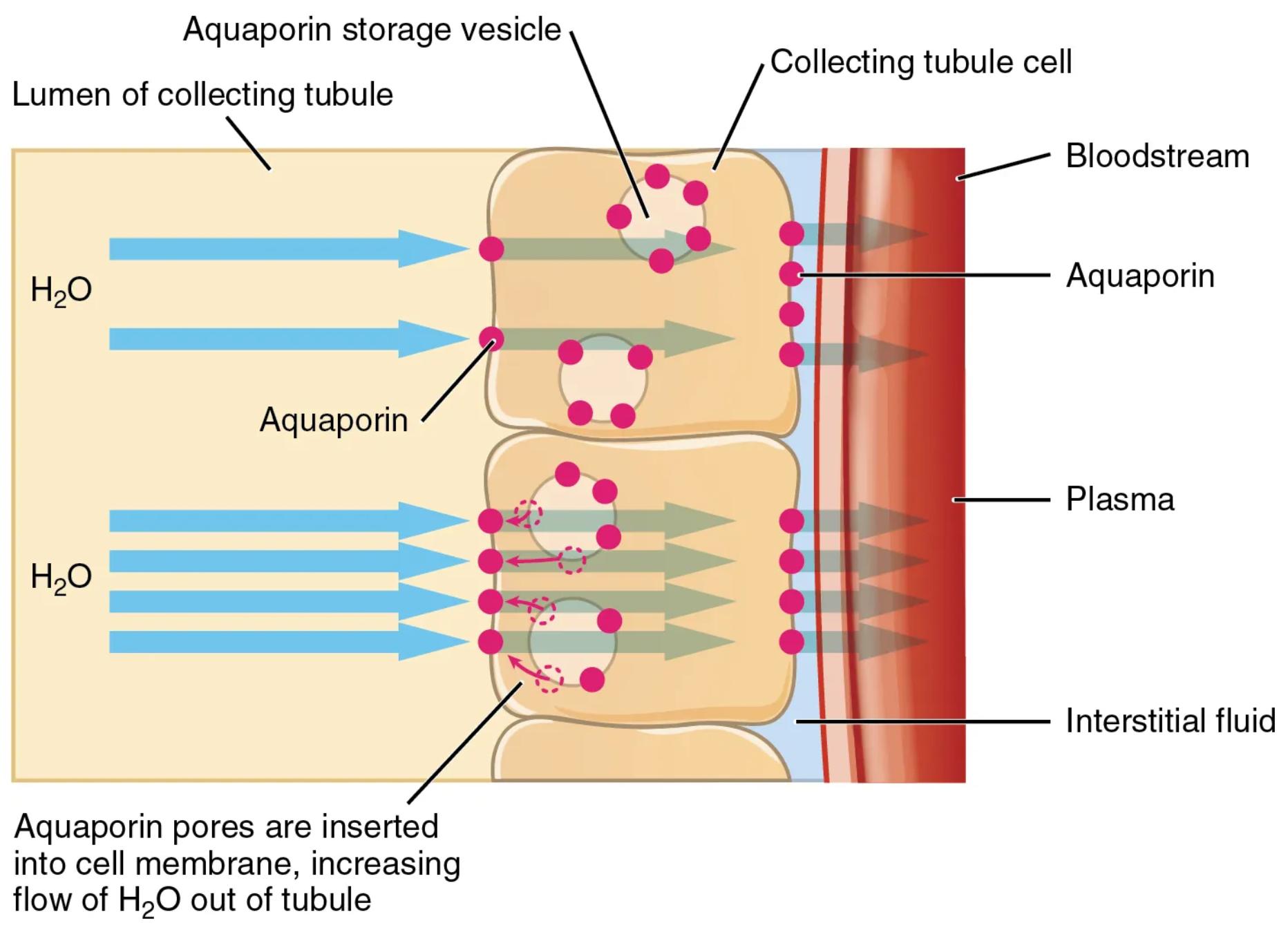
Aquaporin storage vesicle: These are small, membrane-bound sacs within the collecting tubule cells that contain aquaporins. These vesicles act as a reservoir for aquaporins, allowing for rapid insertion into the cell membrane when needed.
Lumen of collecting tubule: This is the inner space or cavity of the collecting tubule, through which the urine filtrate flows. Water and solutes are exchanged between the filtrate in the lumen and the surrounding interstitial fluid.
Collecting tubule cell: These are the epithelial cells that line the collecting tubules in the kidney. They are responsible for regulating the final concentration of urine by reabsorbing water and some solutes.
Bloodstream: This represents the circulating blood within the capillaries adjacent to the collecting tubules. Reabsorbed water from the tubules enters the bloodstream, contributing to the body’s total blood volume.
Aquaporin: These are integral membrane proteins that form pores, or channels, in the cell membrane, specifically designed for the rapid transport of water molecules. They are crucial for highly efficient water movement across biological membranes.
Plasma: This refers to the liquid component of blood, which contains water, proteins, salts, and other substances. Water reabsorbed from the tubules ultimately contributes to the plasma volume.
Interstitial fluid: This is the fluid that surrounds the cells in the tissues, occupying the space between the collecting tubule cells and the bloodstream. Water moves from the tubule lumen, through the collecting tubule cells, into the interstitial fluid, and then into the bloodstream.
Aquaporin pores are inserted into cell membrane, increasing flow of H2O out of tubule: This label describes the key event triggered by ADH. When ADH binds to its receptors on the collecting tubule cells, it signals these cells to move aquaporin-containing vesicles to the apical membrane (facing the lumen) and fuse with it. This insertion dramatically increases the number of water channels, allowing a significant amount of water to flow out of the tubule and into the surrounding interstitial fluid and then the bloodstream.
The Role of Aquaporins in Renal Water Reabsorption
The precise control over water reabsorption in the kidneys is fundamental to maintaining the body’s fluid balance, blood pressure, and overall physiological homeostasis. At the heart of this process are aquaporins, specialized water channels that facilitate the rapid movement of water across cell membranes. This diagram specifically illustrates the mechanism by which aquaporins, under the influence of Antidiuretic Hormone (ADH), enable the collecting tubules to reclaim water from the nascent urine.
In the absence of ADH, the collecting tubule cells have low permeability to water, meaning most water in the filtrate would be excreted. However, when the body needs to conserve water, such as during dehydration, ADH is released. ADH binds to specific receptors on the basolateral membrane of the collecting tubule cells. This binding initiates an intracellular signaling cascade that leads to the translocation of aquaporin-containing vesicles to the apical (luminal) membrane.
-
These vesicles then fuse with the cell membrane, inserting a multitude of aquaporin water channels.
The insertion of these aquaporin pores dramatically increases the cell’s permeability to water. This allows water molecules to rapidly move by osmosis from the lumen of the collecting tubule, where the filtrate is hypotonic relative to the interstitial fluid, through the aquaporin channels, into the collecting tubule cells, and subsequently into the interstitial fluid and finally the bloodstream. This highly efficient reabsorption mechanism is critical for producing concentrated urine and preventing excessive water loss, thereby conserving the body’s vital fluid stores.
Clinical Relevance of Aquaporins
Disruptions in aquaporin function or expression can have significant clinical consequences. For instance, in conditions like nephrogenic diabetes insipidus, the kidneys fail to respond to ADH, often due to defects in the aquaporin-2 gene or the ADH receptor, leading to an inability to reabsorb water and resulting in polyuria (excessive urination) and polydipsia (excessive thirst). Conversely, in conditions where there is excessive water retention, such as in certain edematous states, abnormal regulation of aquaporins can play a role. The detailed understanding of aquaporin dynamics, as depicted here, is therefore crucial for comprehending renal physiology and for the development of therapeutic strategies for a range of fluid balance disorders.
The Microscopic Mechanism of Hydration
The human body’s ability to meticulously manage its water content is a cornerstone of health, and the kidneys are the primary organs responsible for this complex task. Within the nephrons, specifically in the collecting tubules, a sophisticated molecular mechanism involving aquaporins ensures that precisely the right amount of water is reabsorbed from the filtrate back into the bloodstream. This process is not passive; it is tightly regulated, predominantly by Antidiuretic Hormone (ADH), and is vividly illustrated in this detailed diagram.
When the body needs to conserve water, ADH signals the collecting tubule cells to dramatically increase their permeability to water. This occurs through a remarkable cellular event: aquaporin storage vesicles, small intracellular sacs loaded with aquaporin proteins, are mobilized. These vesicles then fuse with the apical plasma membrane of the collecting tubule cells, effectively inserting numerous aquaporin water channels into the membrane facing the lumen of the tubule. This rapid insertion of aquaporin pores transforms the cell membrane from being relatively impermeable to highly permeable to water.
Once these channels are in place, water molecules, or H2O, in the lumen of the collecting tubule can rapidly flow through the aquaporin channels, down their osmotic gradient. This movement carries water out of the tubule cells, through the interstitial fluid, and finally into the bloodstream, where it contributes to maintaining blood volume and pressure. The efficiency of this process is extraordinary, allowing the kidneys to finely tune urine concentration from very dilute to highly concentrated, all in response to the body’s hydration needs. Understanding this intricate cellular machinery provides a profound appreciation for the physiological precision that underpins our body’s ability to maintain fluid balance.

