The red blood cell (RBC) membrane is a masterpiece of biological engineering, designed to withstand the turbulent environment of the circulatory system while maintaining extreme flexibility. Unlike most cells, the erythrocyte must squeeze through capillaries half its diameter and survive hundreds of thousands of cycles of deformation over its 120-day lifespan. This remarkable durability and elasticity are provided by a sophisticated protein network that anchors the lipid bilayer to an underlying cytoskeleton. Understanding these molecular interactions is essential for diagnosing hemolytic anemias and understanding blood group antigens.
Label-by-Label Explanation
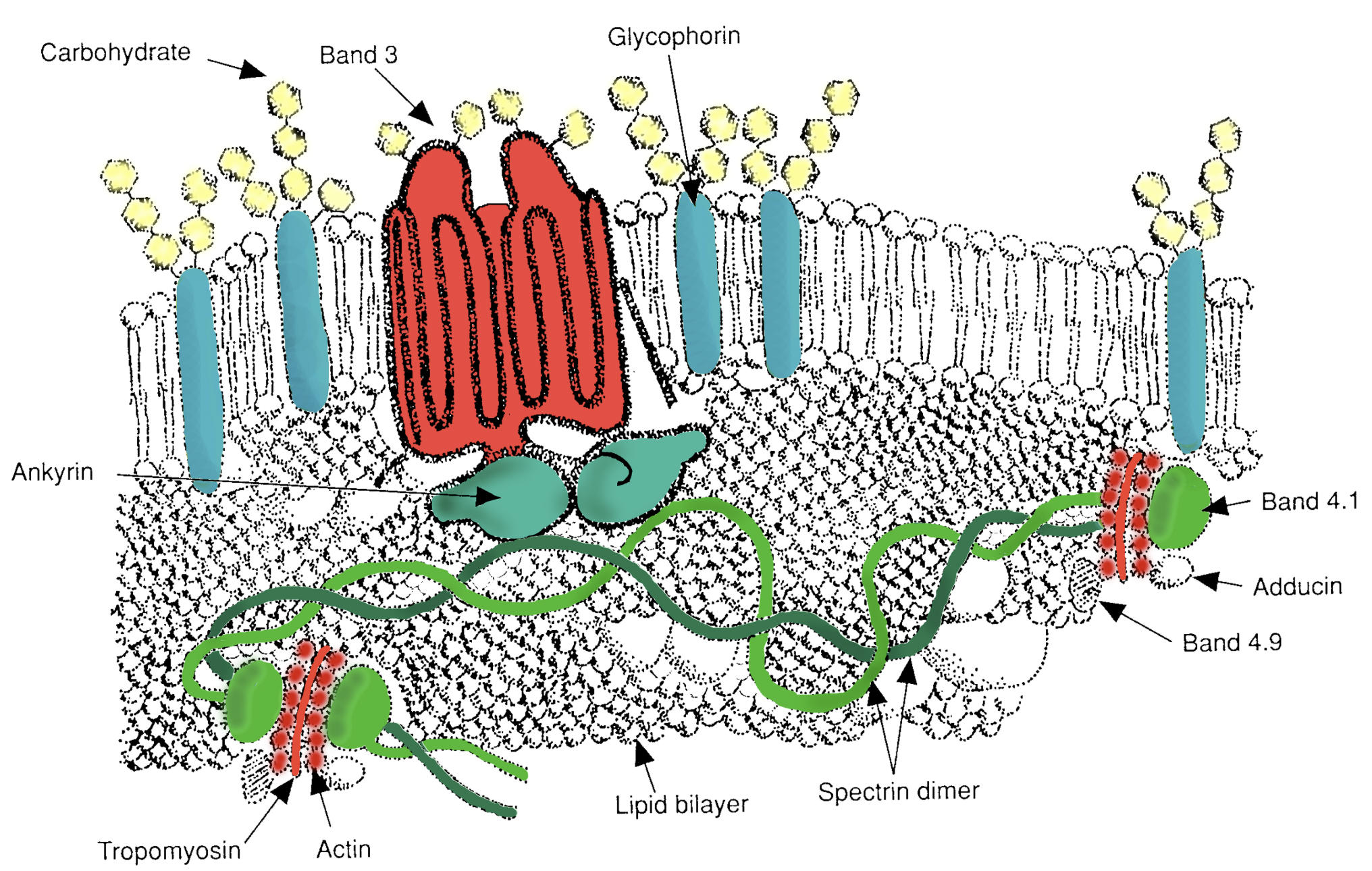
Carbohydrate: These are oligosaccharide chains attached to proteins and lipids on the extracellular surface, forming the glycocalyx. They play a critical role in cell-cell recognition and define the various human blood group systems, such as the ABO system.
Band 3: This is a major multipass transmembrane protein that functions as an anion exchanger, primarily facilitating the exchange of chloride and bicarbonate. It serves as a central anchoring point for the cytoskeleton by binding to ankyrin and other peripheral proteins.
Glycophorin: These are sialoglycoproteins that span the membrane and are heavily glycosylated on their extracellular domain. They contribute to the negative charge of the RBC surface (zeta potential), which prevents red cells from aggregating in the bloodstream.
Ankyrin: This large, globular protein acts as a primary bridge between the transmembrane Band 3 protein and the spectrin cytoskeleton. It is vital for maintaining the vertical stability of the membrane, and its deficiency is a common cause of hereditary spherocytosis.
Band 4.1: This protein stabilizes the “junctional complex” where spectrin filaments meet actin. It significantly enhances the binding affinity of spectrin to actin, ensuring the lateral integrity of the cytoskeletal meshwork.
Adducin: Found at the junctional complexes, this protein promotes the assembly of the spectrin-actin network. It helps cap the fast-growing ends of actin filaments to regulate their length and stability.
Band 4.9: Also known as dematin, this actin-bundling protein is located at the junctional complexes. It helps organize and stabilize the short actin protofilaments that serve as nodes for the spectrin network.
Spectrin dimer: This is the primary structural component of the cytoskeleton, consisting of alpha and beta chains twisted together. Dimers associate head-to-head to form tetramers, creating a flexible, hexagonal lattice that supports the entire lipid bilayer.
Lipid bilayer: This fluid-mosaic structure consists of phospholipids and cholesterol, providing the basic permeability barrier of the cell. It is tethered to the underlying proteins, which prevents the lipid membrane from vesiculating or peeling away during cell deformation.
Tropomyosin: This protein associates with the short actin filaments in the junctional complex. Its role is to stabilize the actin protofilaments, ensuring they maintain the correct length to anchor multiple spectrin molecules.
Actin: Existing as short protofilaments (junctional nodes), actin provides the site where multiple spectrin tetramers converge. This arrangement creates the mesh-like nature of the cytoskeleton, allowing for reversible expansion and contraction.
Functional Significance
The primary function of the RBC membrane protein complex is to provide mechanical stability and reversible deformability. The membrane is organized into two interconnected layers: the fluid lipid bilayer and the elastic protein cytoskeleton. The connections between these layers are often categorized into vertical and horizontal interactions.
- Vertical Interactions: These connect the lipid bilayer to the cytoskeleton, primarily through the Band 3-Ankyrin-Spectrin linkage. These interactions prevent the lipid bilayer from separating from the cell and are essential for maintaining the biconcave shape.
- Horizontal Interactions: These occur within the plane of the cytoskeleton itself, primarily involving the spectrin-actin-protein 4.1 junctional complexes. These interactions provide the tensile strength needed to endure the high shear stress of arterial circulation.
Clinical Relevance
Disruptions in the proteins shown in this image lead to specific clinical pathologies known as hereditary hemolytic anemias. When the vertical linkages (like Ankyrin or Band 3) are weakened, the lipid bilayer loses its support and blebs off as microvesicles. This reduces the cell’s surface-area-to-volume ratio, forcing the cell into a spherical shape (hereditary spherocytosis). These spherocytes are less flexible and are prematurely destroyed by the spleen.
Conversely, defects in horizontal interactions (such as mutations in the spectrin dimer-dimer contact points) lead to mechanical instability of the cytoskeleton. Under shear stress, the cells become permanently deformed and elongated, resulting in hereditary elliptocytosis. In both cases, the fundamental issue is a failure of the molecular architecture to maintain the structural integrity required for normal circulation.
Summary for Medical Learners
The red blood cell membrane is not a static shell but a dynamic, semi-fluid structure. For medical examinations and clinical practice, remember that the spectrin-ankyrin-band 3 axis is the most important vertical tether. Patients presenting with anemia, jaundice, and splenomegaly should be evaluated for defects in these specific proteins. The negative charge provided by glycophorins is equally important, as it keeps RBCs suspended; loss of this charge can contribute to increased blood viscosity and thrombotic risk in certain disease states.
Medical Learning Tips
- Identify ankyrin as the primary vertical bridge connecting the spectrin cytoskeleton to the transmembrane Band 3 protein.
- Recall that defects in horizontal linkages typically cause elliptocytosis, while vertical defects lead to spherocytosis.
- Remember that glycophorins are the primary determinants of the RBC's negative surface charge, which prevents auto-aggregation.

