The arterial system of the thoracic and abdominal regions represents the primary conduit for oxygenated blood distribution from the heart to the rest of the body. Centered around the aorta, the largest artery in the human body, this network is organized into systematic branches that supply the body wall (parietal branches) and the internal organs (visceral branches). Understanding the spatial relationships and branching patterns shown in this anatomical view is fundamental for clinical practice, particularly in vascular surgery, interventional radiology, and emergency medicine, where pathologies such as aneurysms or dissections can have life-threatening consequences.
Label-by-Label Explanation
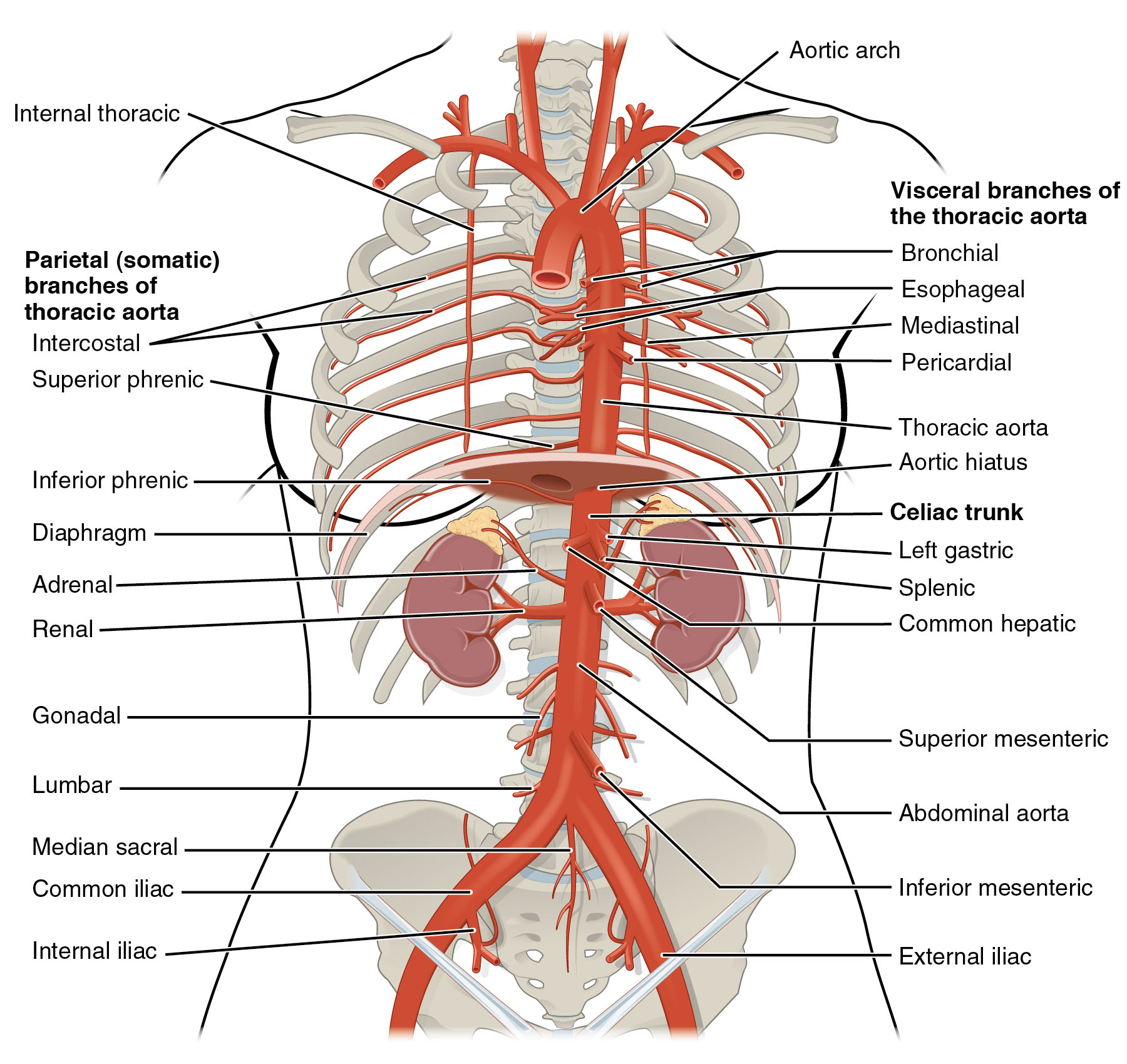
Aortic arch: The curved segment of the aorta that connects the ascending and descending portions; it gives rise to the major vessels supplying the head, neck, and upper limbs.
Internal thoracic: Arising from the subclavian artery, these vessels run along the inner surface of the anterior rib cage to supply the chest wall and mammary glands.
Intercostal: A series of paired parietal branches that travel within the intercostal spaces to provide blood to the ribs, intercostal muscles, and overlying skin.
Superior phrenic: Small parietal branches of the thoracic aorta that supply the superior surface of the diaphragm, assisting in respiratory muscle perfusion.
Bronchial: Visceral branches that provide oxygenated blood to the structural tissues of the lungs and the bronchial tree, distinct from the pulmonary circulation.
Esophageal: Multiple small visceral branches of the thoracic aorta that supply the muscular and mucosal layers of the esophagus during its descent through the mediastinum.
Mediastinal: Small arteries that supply the connective tissue, lymph nodes, and posterior structures within the mediastinal cavity.
Pericardial: Tiny vessels that branch off the thoracic aorta to supply the posterior aspect of the pericardial sac surrounding the heart.
Thoracic aorta: The segment of the descending aorta located within the posterior mediastinum, extending from the aortic arch to the diaphragm.
Aortic hiatus: An opening in the diaphragm at the level of the T12 vertebra that allows the aorta to pass from the thoracic cavity into the abdominal cavity.
Inferior phrenic: The first parietal branches of the abdominal aorta, supplying the inferior surface of the diaphragm and providing branches to the adrenal glands.
Celiac trunk: A major, unpaired visceral branch arising just below the diaphragm that divides into three primary vessels to supply the foregut structures.
Left gastric: The smallest branch of the celiac trunk, which travels along the lesser curvature of the stomach and supplies the lower esophagus.
Splenic: A large, tortuous branch of the celiac trunk that supplies the spleen, parts of the stomach, and the body and tail of the pancreas.
Common hepatic: A branch of the celiac trunk that supplies the liver, gallbladder, duodenum, and parts of the stomach via its various subdivisions.
Superior mesenteric: An unpaired visceral branch arising approximately 1 cm below the celiac trunk, supplying the midgut structures from the duodenum to the transverse colon.
Adrenal: Also known as suprarenal arteries, these paired vessels supply the adrenal glands and may originate directly from the aorta or from the renal/phrenic arteries.
Renal: Large, paired visceral branches that supply the kidneys; they typically arise laterally from the aorta at the level of the L1-L2 vertebrae.
Gonadal: Long, thin paired vessels (testicular in males, ovarian in females) that descend through the retroperitoneum to supply the reproductive organs.
Lumbar: Usually four pairs of parietal branches that supply the posterior abdominal wall, spinal cord, and meninges.
Abdominal aorta: The segment of the descending aorta that begins after passing through the diaphragm and ends at the level of L4 where it bifurcates.
Inferior mesenteric: An unpaired visceral branch arising near the L3 level that supplies the hindgut structures, including the distal colon and rectum.
Median sacral: A small, unpaired artery arising from the posterior aspect of the aortic bifurcation that supplies the lower lumbar vertebrae and sacrum.
Common iliac: The two terminal branches of the abdominal aorta that diverge to supply the pelvis and lower extremities.
Internal iliac: The medial branch of the common iliac artery that supplies the pelvic viscera, perineum, and gluteal region.
External iliac: The lateral branch of the common iliac artery that continues toward the thigh, becoming the femoral artery once it passes the inguinal ligament.
Anatomical Overview
The arterial supply illustrated here demonstrates a highly organized hierarchy. The thoracic aorta is primarily responsible for supplying the thoracic wall and the viscera of the mediastinum. Once the vessel traverses the aortic hiatus at the T12 vertebral level, it becomes the abdominal aorta. The abdominal segment is characterized by three distinct types of branches: unpaired visceral branches (celiac trunk, SMA, IMA), paired visceral branches (renal, adrenal, gonadal), and parietal branches (lumbar, phrenic).
The transition between the superior mesenteric and inferior mesenteric arteries marks the physiological transition from the midgut to the hindgut. Spatially, the aorta lies slightly to the left of the midline and sits anterior to the vertebral column, which serves as a vital landmark during surgical procedures and radiologic assessments.
Clinical Relevance
Knowledge of these branches is essential for diagnosing and managing several vascular conditions. Abdominal Aortic Aneurysm (AAA) often occurs below the level of the renal arteries; understanding the distance between the renal ostia and the aneurysm is critical for determining if a patient is a candidate for endovascular repair (EVAR). Furthermore, the celiac trunk and mesenteric arteries are prone to atherosclerotic narrowing, which can lead to chronic mesenteric ischemia, presenting as “abdominal angina” or post-prandial pain.
In the thoracic region, the origin of the intercostal arteries is a significant consideration during thoracic surgery or chest tube placement. Clinicians must also be aware of the aortic dissection, where a tear in the intimal layer creates a false lumen that can shear off or occlude any of the major branches labeled in the image, leading to organ infarction (e.g., renal failure if the renal arteries are involved or limb ischemia if the iliacs are compromised).
Educational Importance for Medical Learners
For students, mastering the vertebral levels of these branches is a common requirement. The celiac trunk usually appears at T12, the superior mesenteric artery at L1, and the inferior mesenteric artery at L3. The bifurcation into the common iliac arteries typically occurs at the L4 level. Visualizing these vessels in relation to organs like the kidneys and the diaphragm helps in interpreting cross-sectional imaging such as CT scans.
Additionally, understanding the collateral circulation is vital. For example, if the inferior mesenteric artery is occluded, the marginal artery of Drummond can often provide collateral flow from the superior mesenteric artery to the distal colon. This anatomical redundancy highlights the body’s protective mechanisms against focal vascular compromise.
Key Learning Points
- The aorta is divided into thoracic and abdominal segments by the diaphragm at the T12 level.
- Visceral branches supply organs, while parietal (somatic) branches supply the body wall and musculoskeletal structures.
- The three major unpaired visceral branches of the abdominal aorta are the celiac trunk, superior mesenteric artery, and inferior mesenteric artery.
- The renal arteries are large paired branches that typically arise just below the level of the superior mesenteric artery.
- The common iliac arteries represent the terminal bifurcation of the abdominal aorta at the L4 vertebral level.
Medical Learning Tips
- Use the mnemonic 'C-S-I' to remember the order of unpaired visceral branches: Celiac, Superior mesenteric, Inferior mesenteric.
- The aortic bifurcation occurs at the L4 level, which corresponds externally to the level of the iliac crests.
- Always distinguish between the pulmonary circulation and the bronchial arteries; the latter provides the actual nutritional supply to lung tissue.

