Vibrio bacteria represent a distinct genus of Gram-negative bacteria characterized by their unique curved-rod or comma-shaped morphology. Primarily found in salty or brackish water, these microorganisms are of significant clinical interest due to their role in severe gastrointestinal diseases and wound infections. This guide examines the structural anatomy of the vibrio cell and details the pathogenesis of its most notorious member, Vibrio cholerae.
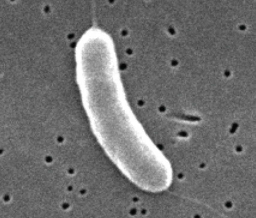
Cell body: The main body of a vibrio is shaped like a curved rod, which provides an efficient surface-area-to-volume ratio for nutrient absorption. This comma-like structure is stabilized by the peptidoglycan layer and specific proteins that guide the cell’s growth into its characteristic curve.
Flagellum: A single, polar flagellum is present at one end of the cell, providing the bacterium with high-speed motility. This whip-like appendage allows the vibrio to navigate through viscous environments, such as the mucus lining of the human intestine, to establish an infection.
The genus Vibrio consists of facultative anaerobes that are mostly halophilic, meaning they require or prefer saline environments for optimal growth. These bacteria are ubiquitous in aquatic ecosystems, where they often attach to the chitinous shells of copepods and other shellfish. While many species are harmless, others are potent human pathogens that can cause rapid-onset illness when ingested through contaminated food or water.
The architectural simplicity of the vibrio cell belies its complex survival strategies. From its high metabolic rate to its ability to enter a “viable but non-culturable” (VBNC) state under environmental stress, this organism is built for resilience. In clinical settings, the rapid identification of vibrio species is crucial, as some infections can lead to life-threatening dehydration within hours.
Key biological and ecological characteristics of Vibrio include:
- Facultative anaerobic metabolism, allowing survival in both oxygen-rich and oxygen-poor environments.
- A characteristic curved-rod (comma) shape that aids in movement and identification.
- Possession of a single, polar flagellum for rapid motility and chemotaxis.
- Presence of two circular chromosomes, a unique genetic feature among many bacterial genera.
Pathogenesis and the Clinical Presentation of Cholera
The most significant medical threat posed by this genus is cholera, an acute diarrheal infection caused by the ingestion of food or water contaminated with the bacterium Vibrio cholerae. Once inside the host, the bacteria use their motility to reach the small intestine, where they adhere to the mucosal surface. They do not typically invade the intestinal wall; instead, they produce a potent enterotoxin known as cholera toxin.
This toxin disrupts the normal physiology of the intestinal epithelial cells by increasing intracellular levels of cyclic AMP. This biochemical shift leads to a massive secretion of water and electrolytes, particularly chloride and sodium, into the intestinal lumen. The result is the hallmark symptom of cholera: voluminous “rice-water” stools that can lead to severe dehydration and hypovolemic shock if left untreated.
Management of cholera focuses primarily on aggressive rehydration therapy, which involves the administration of oral rehydration salts (ORS) or intravenous fluids to replace lost volume and electrolytes. While most cases can be managed with rehydration alone, antibiotics may be used to reduce the duration of the illness and the shedding of bacteria. Prevention remains the most effective strategy, centered on improving water sanitation, hygiene practices, and the use of oral cholera vaccines in endemic areas.
Anatomical Protection and Environmental Resilience
Beyond cholera, other vibrio species like Vibrio vulnificus and Vibrio parahaemolyticus are known for causing severe wound infections and foodborne gastroenteritis, often associated with consuming raw or undercooked seafood. These bacteria are Gram-negative, meaning they possess a thin peptidoglycan layer surrounded by an outer membrane containing lipopolysaccharides (LPS). This outer membrane acts as an additional protective barrier against certain antibiotics and host immune defenses.
The study of vibrio bacteria bridges the gap between environmental microbiology and clinical medicine. Their unique comma-shaped anatomy and specialized motility mechanisms make them highly successful survivors in both natural oceans and human hosts. By maintaining rigorous public health standards and continuing to research the molecular mechanisms of vibrio pathogenesis, we can better protect global populations from the devastating impacts of waterborne diseases.

