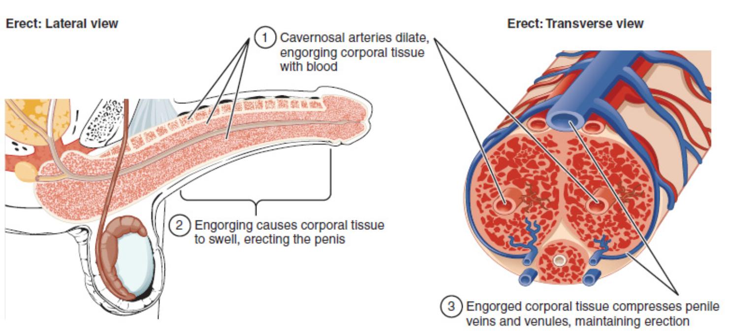
This article provides a comprehensive overview of the physiological mechanisms involved in achieving and maintaining an erection, as illustrated by the provided lateral and transverse views of the erect penis. We will explore the crucial steps from arterial dilation and corporal tissue engorgement to the venous compression that sustains rigidity. Understanding this intricate process is fundamental to comprehending male sexual function and related health conditions.
Explaining the Stages of Erection
1 Cavernosal arteries dilate, engorging corporal tissue with blood: During sexual arousal, parasympathetic nerve stimulation causes the release of nitric oxide, leading to the relaxation of smooth muscle in the walls of the cavernosal arteries. This vasodilation significantly increases blood flow into the corpora cavernosa and corpus spongiosum, filling the sinusoidal spaces.
2 Engorging causes corporal tissue to swell, erecting the penis: As blood rapidly fills the vascular spaces within the corpora cavernosa, the erectile tissue expands and hardens. This volumetric expansion is the primary physical event that causes the penis to become erect, providing the necessary rigidity for intercourse.
3 Engorged corporal tissue compresses penile veins and venules, maintaining erection: The expansion of the corpora cavernosa against the tunica albuginea (a dense fibrous capsule surrounding the erectile tissue) leads to the compression of the subtunical venules. This veno-occlusion mechanism significantly reduces venous outflow from the penis, effectively trapping blood within the erectile chambers and sustaining the erection.
The process of penile erection is a complex neurovascular event, meticulously orchestrated by the nervous system and vascular responses. It represents a dramatic shift from the flaccid state, transforming the penis into a rigid structure capable of sexual intercourse. This dynamic transformation is critical for reproductive success and is a hallmark of male sexual health.
At its core, an erection is a hydraulic event. The increase in arterial blood flow into the erectile tissues, primarily the corpora cavernosa, is the initial and most vital step. This influx of blood is not a passive process but an actively regulated one, responding to various stimuli that trigger the relaxation of smooth muscles within the penile vasculature.
The efficiency of this system is remarkable, ensuring that once sufficient blood has entered the erectile chambers, it is effectively retained. This retention is achieved through a sophisticated veno-occlusive mechanism, where the expanding erectile tissue compresses the outflow veins. Disruptions to any part of this intricate cascade can lead to erectile dysfunction, highlighting the delicate balance required for normal penile function. Understanding these mechanisms is crucial for appreciating the physiological basis of male sexual response.
The intricate dance between arterial inflow and venous outflow is at the heart of erectile physiology. When the cavernosal arteries dilate, blood rushes into the erectile bodies, causing them to engorge. This engorgement is not merely a swelling; it is a structural change that results in increased rigidity. The subsequent compression of the penile veins by the expanding corporal tissue serves as a biological one-way valve, preventing blood from leaving the penis too quickly and thereby sustaining the erection. This veno-occlusion mechanism is a vital component in maintaining penile rigidity for the duration of sexual activity. Issues such as veno-occlusive dysfunction, where the veins fail to compress adequately, can lead to the inability to maintain an erection. Therefore, a comprehensive understanding of these processes is essential for diagnosing and treating various forms of erectile dysfunction.
Conclusion
The detailed explanation of the erect penis, encompassing both lateral and transverse views, illuminates the sophisticated physiological processes involved in achieving and maintaining an erection. From the initial arterial dilation and engorgement of corporal tissues to the critical veno-occlusion mechanism, each step is vital. This deep understanding of penile physiology is fundamental not only for appreciating normal male sexual function but also for diagnosing and treating conditions that impair this essential aspect of reproductive health.

