The right hip joint is a marvel of human anatomy, serving as a ball-and-socket joint that supports the body’s weight and enables a wide range of movements. This detailed illustration, showcasing frontal, anterior, and posterior views, highlights the bones, ligaments, and cartilage that work together to ensure stability and flexibility. Understanding these structures provides valuable insight into how the hip functions in daily activities and maintains overall lower body health.
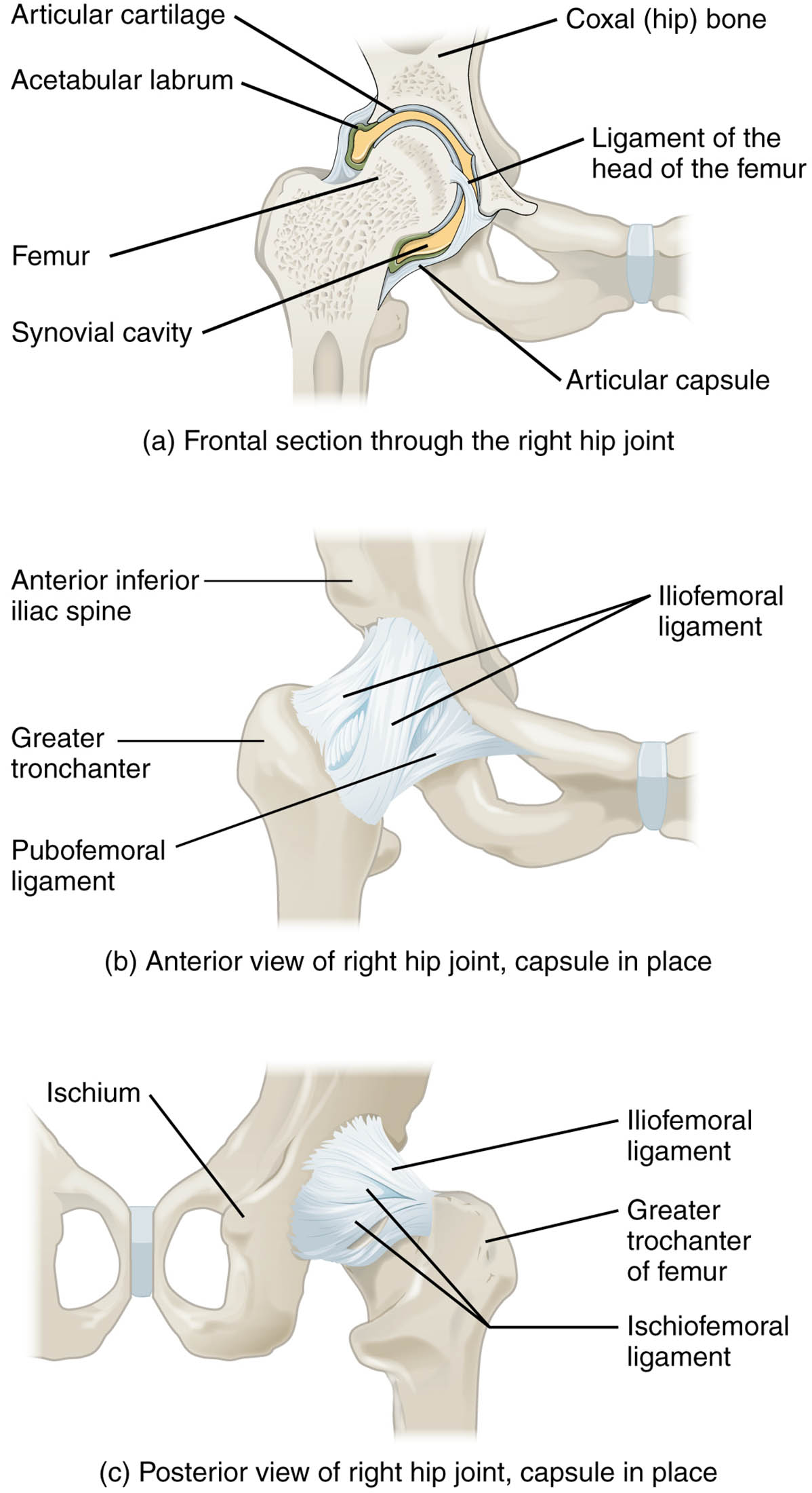
Labeled Parts Explanation
- Articular cartilage: This smooth, slippery tissue covers the surfaces of the femur and coxal bone, reducing friction and absorbing shock during movement. It plays a crucial role in maintaining joint health by preventing bone-on-bone contact.
- Acetabular labrum: This ring of cartilage surrounds the acetabulum, deepening the socket to enhance stability and secure the femoral head. It also helps distribute weight and improve the joint’s load-bearing capacity.
- Ligament of the head of the femur: This small ligament connects the femoral head to the acetabulum, providing additional stability and carrying blood vessels that nourish the bone. Though less strong than other ligaments, it contributes to joint integrity.
- Femur: The thigh bone forms the ball of the hip joint, articulating with the acetabulum to allow multiaxial movement. Its strength and structure are essential for weight-bearing and locomotion.
- Synovial cavity: This space within the joint contains synovial fluid, which lubricates the articular surfaces and nourishes the cartilage. It ensures smooth movement and reduces wear on the joint components.
- Articular capsule: This fibrous structure encloses the hip joint, providing a protective barrier and limiting excessive motion. It contains the synovial cavity and supports the joint’s stability.
- Coxal (hip) bone: This large pelvic bone forms the socket of the hip joint, known as the acetabulum, and anchors the femur. It plays a key role in weight distribution and pelvic stability.
- Anterior inferior iliac spine: This bony projection on the coxal bone serves as an attachment point for ligaments and muscles, aiding in hip flexion. It contributes to the overall structural support of the pelvis.
- Iliofemoral ligament: One of the strongest ligaments, it connects the ilium to the femur, preventing hyperextension of the hip. It tightens during standing, pulling the femoral head into the acetabulum.
- Greater trochanter: This prominent bony protrusion on the femur serves as an attachment site for major hip muscles like the gluteus medius. It facilitates abduction and rotation of the hip joint.
- Pubofemoral ligament: This ligament extends from the pubis to the femur, limiting excessive abduction and extension. It works with other ligaments to maintain hip stability during movement.
- Ischium: This posterior part of the coxal bone forms the lower boundary of the acetabulum and supports weight when sitting. It also provides attachment points for ligaments and muscles.
- Ischiofemoral ligament: This ligament connects the ischium to the femur, restricting internal rotation and hyperextension. It complements the iliofemoral ligament in stabilizing the posterior hip.
Introduction to Hip Joint Anatomy
The hip joint is a critical component of the lower body, functioning as a ball-and-socket joint that supports mobility and stability. This illustration offers a detailed look at the right hip from multiple angles, revealing the interplay of bones like the femur and coxal bone with supporting ligaments. Grasping these anatomical features enhances your understanding of how the hip withstands daily stresses and facilitates movements such as walking or running.
- Offers a clear view of the hip’s structural complexity.
- Emphasizes the joint’s role in weight-bearing and locomotion.
Ligament Support and Joint Stability
Ligaments are vital for holding the hip joint together, with each playing a specific role. The iliofemoral ligament, known for its strength, prevents the hip from overextending, while the pubofemoral ligament limits abduction. The ischiofemoral ligament adds posterior support, ensuring the joint remains secure during various motions.
- Details how ligaments work together to protect the joint.
- Highlights the synovial cavity’s role in lubrication and nourishment.
Bone Structure and Cartilage Function
The hip’s bony framework includes the femur, with its greater trochanter, and the coxal bone, featuring the ischium and anterior inferior iliac spine. The acetabular labrum and articular cartilage enhance the joint’s surface, providing cushioning and depth to the socket. These elements ensure the hip can handle significant loads while allowing fluid movement.
- Explains the femur’s contribution to hip mobility.
- Describes how cartilage prevents joint degeneration.
Clinical Insights and Functional Importance
Understanding hip anatomy is key to recognizing potential issues, such as ligament strains or cartilage wear. The articular capsule and ligaments like the ligament of the head of the femur are critical in maintaining joint alignment, especially under stress. This knowledge can guide therapeutic approaches to restore function after injury or surgery.
- Provides context for common hip-related conditions.
- Stresses the importance of anatomical awareness in treatment.
Conclusion
The right hip joint’s anatomy, as depicted in this illustration, showcases a harmonious balance of bones, ligaments, and cartilage that supports the body’s movement and stability. From the robust iliofemoral ligament to the protective acetabular labrum, each structure contributes to a resilient joint capable of enduring daily demands. Exploring this anatomy not only deepens your appreciation of the human body but also equips you with the insight to address hip health effectively.

