The venous system of the lower limbs is a vital component of the circulatory network, responsible for returning deoxygenated blood from the legs and feet back to the heart. This anterior view highlights the intricate pathways of major veins, supported by one-way valves and muscular contractions that combat gravity to maintain efficient blood flow. Exploring these structures offers valuable insights into their role in supporting mobility and preventing circulatory challenges.
Detailed Anatomy of Labeled Veins
The following sections provide a comprehensive overview of each labeled vein, detailing their anatomical paths and functional significance.
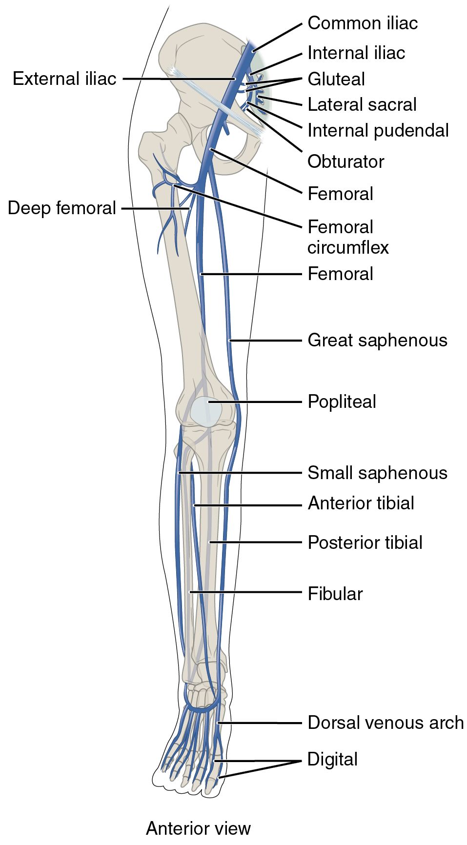
Common iliac: The common iliac vein forms at the pelvic brim where the external and internal iliac veins converge, serving as a primary route for blood from the lower limbs and pelvis. It ascends to join the opposite common iliac vein, forming the inferior vena cava that delivers blood to the heart.
Internal iliac: The internal iliac vein collects blood from the pelvic organs, gluteal region, and internal genitalia, merging with the external iliac to create the common iliac. Its extensive network of tributaries ensures efficient drainage from the pelvic cavity and lower spine.
Gluteal: The gluteal veins drain the gluteal muscles and surrounding tissues, running alongside the gluteal arteries to join the internal iliac vein. These veins are essential for maintaining circulation in the buttocks, supporting posture and movement.
Lateral sacral: The lateral sacral veins gather blood from the sacral region and vertebral column, feeding into the internal iliac vein. They play a critical role in preventing venous pooling in the lower back and pelvic area.
Internal pudendal: The internal pudendal vein drains the perineal region, including the external genitalia and anal area, following the pudendal artery’s path. It contributes to the internal iliac vein, aiding venous return from the pelvic floor.
Obturator: The obturator vein drains the adductor muscles and hip joint, passing through the obturator canal to join the internal iliac vein. This vessel supports circulation in the medial thigh, facilitating hip stability.
Femoral: The femoral vein is a key deep vein that begins as the popliteal vein and runs through the thigh, transitioning into the external iliac at the inguinal ligament. It receives blood from both deep and superficial sources, playing a central role in lower limb venous return.
Femoral circumflex: The femoral circumflex veins, including medial and lateral branches, drain the upper thigh and hip region, accompanying the circumflex arteries. They typically empty into the femoral or deep femoral veins, supporting circulation around the hip joint.
Deep femoral: The deep femoral vein, or profunda femoris, runs parallel to the deep femoral artery, draining the deeper thigh muscles. It joins the femoral vein, providing a vital pathway for blood from the quadriceps and hamstrings.
Great saphenous: The great saphenous vein, the longest superficial vein, originates from the dorsal venous arch and ascends medially to join the femoral vein at the saphenous opening. It drains the medial and anterior leg and thigh, often utilized in vascular surgeries.
Popliteal: The popliteal vein forms behind the knee from the union of the anterior and posterior tibial veins, continuing as the femoral vein. It drains the knee joint and calf muscles, relying on valves to prevent backflow during movement.
Small saphenous: The small saphenous vein runs along the posterior leg from the lateral foot to the popliteal fossa, joining the popliteal vein. It drains the lateral and posterior leg, complementing the great saphenous in superficial circulation.
Anterior tibial: The anterior tibial vein drains the anterior compartment of the leg, including dorsiflexor muscles, and ascends to form the popliteal with the posterior tibial. Paired with its artery, it ensures blood return from the shin critical for foot movement.
Posterior tibial: The posterior tibial vein runs deep in the calf, draining the plantar foot and posterior leg muscles, joining the anterior tibial at the popliteal. It supports upward blood flow through calf muscle action and perforating veins.
Fibular: The fibular vein, or peroneal, accompanies the fibular artery in the lateral calf, draining deep tissues and merging with the posterior tibial. This vein aids circulation in the lateral leg, supporting balance and peroneal muscle function.
Dorsal venous arch: The dorsal venous arch is a network on the foot’s dorsum, collecting blood from the toes and feeding into the great and small saphenous veins. It serves as the foundation for superficial venous return in the lower limb.
Digital: The digital veins drain the toes, forming arcs on the dorsal and plantar sides of the foot. They connect to the dorsal venous arch, ensuring efficient clearance of deoxygenated blood from the distal extremities.
Physiological Role of Lower Limb Veins
The veins of the lower limbs are engineered to overcome gravitational challenges, relying on muscular pumps and valves for effective blood return.
- Deep veins like the femoral and popliteal handle the majority of venous blood, benefiting from the skeletal muscle pump during walking or exercise.
- Superficial veins such as the great and small saphenous provide an auxiliary route, connecting to deep veins via perforators for heat regulation.
- Valves ensure unidirectional flow, preventing reflux and maintaining pressure gradients toward the heart.
- Blood from the foot ascends through the dorsal arch and tibial veins, merging at key points to enter the thigh and pelvis seamlessly.
Clinical Relevance and Maintenance
Knowledge of venous anatomy aids in managing common circulatory issues, promoting proactive health strategies.
- Varicose veins often involve the great saphenous, resulting from valve incompetence and increased pressure, leading to visible bulging.
- Deep vein thrombosis may affect the femoral or popliteal veins, where clots from immobility pose a risk of pulmonary embolism.
- Treatments like compression stockings or vein ablation target problematic veins while preserving deep circulation.
- Regular movement and leg elevation enhance venous return, mimicking the natural pump to reduce swelling.
Connection to Systemic Circulation
The lower limb veins integrate with the broader circulatory system, supporting overall physiological balance.
- Blood from the common iliac joins the inferior vena cava, delivering it to the right atrium for oxygenation.
- Hormones like aldosterone from the adrenal glands influence vascular tone, affecting fluid dynamics in these veins.
- Prolonged standing or pregnancy can increase pressure on iliac veins, causing temporary edema that movement can alleviate.
- Lymphatic vessels alongside these veins manage interstitial fluid, complementing venous drainage and immune function.
In conclusion, the major veins of the lower limbs form a resilient network that adapts to the demands of movement and posture. A deeper understanding of their anatomy encourages habits that sustain healthy circulation, benefiting long-term vascular health.

