Capillaries are the smallest blood vessels in the circulatory system, serving as critical sites for the exchange of oxygen, nutrients, and waste between blood and tissues. This image provides a detailed view of the continuous type of capillary, showcasing its unique structure that supports various physiological processes across different organs.
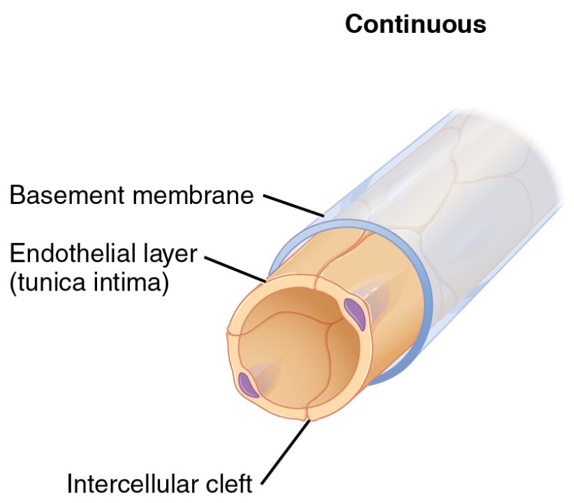
Basement membrane The basement membrane is a thin, supportive layer beneath the endothelial cells, providing structural stability to the capillary wall. It acts as a selective filter, allowing small molecules like glucose to pass while restricting larger substances to maintain tissue integrity.
Endothelial layer (tunica intima) The endothelial layer, or tunica intima, comprises a single layer of flattened cells that line the interior of the capillary. This layer facilitates the exchange of gases and nutrients, while also playing a key role in preventing blood clotting and regulating vascular tone.
Intercellular cleft The intercellular cleft represents the small gaps between adjacent endothelial cells in continuous capillaries. These spaces permit the controlled diffusion of water, ions, and small solutes, ensuring efficient exchange without compromising the barrier function.
The Role of Continuous Capillaries in the Body
Continuous capillaries are essential for maintaining a stable environment in various tissues. Their tightly structured design makes them ideal for regions requiring controlled substance exchange.
- They form the foundation of the blood-brain barrier, protecting the central nervous system by limiting the passage of harmful substances.
- Found abundantly in muscles, skin, and the lungs, they support steady oxygen and nutrient delivery to active tissues.
- The complete basement membrane enhances their role as a selective filter, crucial for homeostasis.
- In the lungs, they facilitate the exchange of oxygen and carbon dioxide, aligning with respiratory needs.
Anatomical Features of Continuous Capillaries
The structure of continuous capillaries is defined by their endothelial and supportive layers. These features adapt to the specific demands of the tissues they serve.
- The endothelial layer is tightly joined, with minimal gaps to prevent leakage of plasma proteins.
- Basement membrane surrounds the endothelium, offering additional support and filtration.
- Intercellular clefts are narrow, allowing only small molecules to pass through via diffusion.
- Their uniform structure contrasts with other capillary types, making them suited for protective roles.
Physiological Functions and Importance
Continuous capillaries play a vital role in sustaining tissue health through regulated exchange processes. Their design ensures that critical substances reach their targets without excessive fluid loss.
- They support the delivery of hormones like T3 and T4 from the thyroid gland to regulate metabolism.
- In skeletal muscles, they provide oxygen during physical activity, aiding energy production.
- The tight junctions prevent edema by minimizing fluid leakage into interstitial spaces.
- Their presence in the nervous system underscores their protective function against toxins.
Clinical Significance of Continuous Capillaries
Understanding the anatomy of continuous capillaries can shed light on various health conditions. Changes in their structure or function may indicate underlying issues that require attention.
- Damage to the endothelial layer can contribute to inflammation or atherosclerosis in vascular diseases.
- In diabetes, altered permeability may lead to microvascular complications affecting the eyes and kidneys.
- The basement membrane can thicken in chronic conditions, impacting nutrient exchange.
- Research into capillary health supports advancements in treatments for circulatory disorders.
Comparison with Other Capillary Types
While continuous capillaries offer a tight barrier, other types like fenestrated and sinusoid capillaries serve different purposes. This comparison highlights their unique adaptations within the vascular system.
- Unlike fenestrated capillaries, continuous types lack pores, making them less permeable.
- Sinusoid capillaries have wider gaps and incomplete membranes, contrasting with the tight structure here.
- The intercellular cleft in continuous capillaries is smaller than the gaps in sinusoids, limiting cell passage.
- This distinction ensures each type meets the specific needs of organs like the liver or kidneys.
Maintenance and Regulation of Continuous Capillaries
The body maintains continuous capillaries through a balance of cellular and molecular mechanisms. These processes ensure their longevity and functionality in diverse physiological contexts.
- Endothelial cells release nitric oxide to regulate blood flow and prevent clotting.
- The basement membrane is continuously remodeled to adapt to tissue demands.
- Local factors like pH and oxygen levels influence capillary dilation or constriction.
- This regulation supports efficient exchange during rest or increased metabolic activity.
In conclusion, the continuous capillary structure, as depicted in this image, exemplifies the body’s remarkable ability to tailor its vascular network to specific needs. With its intact basement membrane, cohesive endothelial layer, and controlled intercellular clefts, this capillary type ensures precise substance exchange while safeguarding delicate tissues. Exploring these features deepens our appreciation of the circulatory system’s complexity and its critical role in maintaining health.

