
Capillaries are the microscopic vessels that facilitate the exchange of substances between blood and tissues, and fenestrated capillaries stand out due to their specialized structure. This image highlights the fenestrated type of capillary, revealing its unique features that enhance permeability and support critical physiological processes in specific organs.
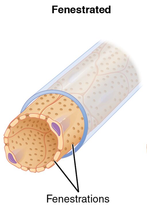
Fenestrations Fenestrations are small, round pores or openings within the endothelial cells of fenestrated capillaries. These pores, typically 60-80 nanometers in diameter, allow for rapid exchange of fluids, solutes, and small molecules, making them essential for filtration and absorption in certain tissues.
The Role of Fenestrated Capillaries in the Body
Fenestrated capillaries play a pivotal role in organs requiring high exchange rates. Their porous structure supports functions critical to maintaining bodily homeostasis.
- They are predominantly found in the kidneys, where they aid in the filtration of blood to form urine.
- In the endocrine glands, such as the pancreas, they facilitate the release of hormones like insulin into the bloodstream.
- The small fenestrations enhance the uptake of nutrients in the intestinal villi during digestion.
- Their design supports rapid fluid movement, aligning with the needs of high-metabolic-rate tissues.
Anatomical Structure of Fenestrated Capillaries
The anatomy of fenestrated capillaries is tailored to their function, featuring a distinctive endothelial layer. This structure sets them apart from other capillary types like continuous or sinusoid.
- The endothelial cells contain numerous fenestrations, which are evenly distributed across the surface.
- A continuous basement membrane underlies the endothelium, providing structural support while allowing selective permeability.
- These capillaries lack the tight junctions seen in continuous types, promoting greater exchange.
- The pores are covered by a thin diaphragm in some cases, regulating the size of molecules that pass through.
Physiological Functions and Importance
The presence of fenestrations enables fenestrated capillaries to handle specialized tasks efficiently. This adaptability is crucial for the health of organs involved in secretion and filtration.
- In the kidneys, they filter plasma to remove waste, contributing to the production of urine.
- Endocrine organs use them to secrete hormones like T3 and T4 from the thyroid gland into circulation.
- They support nutrient absorption in the small intestine, aiding digestion and metabolism.
- The controlled permeability helps maintain fluid balance and prevent excessive leakage.
Clinical Significance of Fenestrated Capillaries
Understanding the structure and function of fenestrated capillaries can provide insights into various medical conditions. Alterations in their permeability may signal underlying health issues.
- Damage to fenestrations can lead to proteinuria, where proteins leak into urine due to kidney dysfunction.
- In diabetes, these capillaries may experience thickening of the basement membrane, impairing filtration.
- They are targeted in drug delivery systems for conditions like cancer, leveraging their permeability.
- Research into their repair mechanisms could improve treatments for renal and endocrine disorders.
Comparison with Other Capillary Types
Fenestrated capillaries differ from continuous and sinusoid types, each serving unique purposes in the vascular network. This comparison highlights their specialized role within the body.
- Unlike continuous capillaries, fenestrated types have fenestrations that increase permeability.
- Sinusoid capillaries feature larger gaps and incomplete membranes, contrasting with the controlled pores here.
- The basement membrane remains intact in fenestrated capillaries, unlike the discontinuous structure in sinusoids.
- This distinction allows fenestrated capillaries to excel in filtration and secretion tasks.
Maintenance and Regulation of Fenestrated Capillaries
The body regulates fenestrated capillaries to ensure they function optimally under varying conditions. These mechanisms support their role in dynamic physiological processes.
- Endothelial cells adjust the size of fenestrations in response to hormonal signals.
- The basement membrane is maintained to balance support and permeability.
- Blood flow and pressure influence the efficiency of exchange through these pores.
- Local factors like inflammation can alter their function, affecting organ performance.
In conclusion, the fenestrated capillary, as depicted in this image, showcases a remarkable adaptation for enhanced exchange within the circulatory system. With its characteristic fenestrations, this capillary type supports vital functions in the kidneys, endocrine glands, and intestines, ensuring efficient filtration and secretion. Exploring these features deepens our understanding of how the body maintains its intricate balance and responds to physiological demands.


