The anatomy of blood vessels is a cornerstone of the circulatory system, with their sectional views revealing the intricate layers that facilitate blood transport throughout the body. This image provides a detailed look at the tunica intima, tunica media, and tunica adventitia, showcasing the structural adaptations that support the high-pressure demands of arteries and the lower-pressure needs of veins.
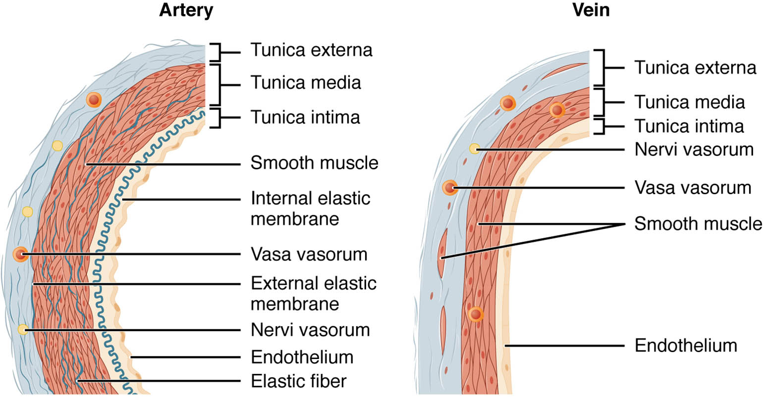
Tunica intima Tunica intima is the innermost layer of blood vessels, consisting of a single layer of endothelial cells that minimize friction and promote smooth blood flow. This layer also contains a subendothelial layer and, in arteries, an internal elastic lamina, which enhances its resilience under pressure.
Tunica media Tunica media forms the middle layer, composed of smooth muscle cells and elastic fibers that regulate vessel diameter and withstand blood pressure. In arteries, this layer is thicker to accommodate high pressure, while in veins, it is thinner, reflecting their role in lower-pressure environments.
Tunica adventitia Tunica adventitia is the outermost layer, made up of connective tissue and collagen fibers that anchor the vessel to surrounding structures. This layer provides structural support and contains vasa vasorum, small vessels that nourish the larger vessel walls, particularly in thicker arteries.
The Role of Layered Structure in Blood Vessels
This exploration reveals how each layer contributes to the vessel’s function. The distinct composition of these layers ensures efficient blood circulation tailored to specific needs.
- Tunica Intima Function: The endothelial lining prevents clot formation by releasing nitric oxide, a vasodilator. This layer’s smoothness reduces turbulence, protecting against vascular damage.
- Tunica Media Role: The smooth muscle in this layer allows vasoconstriction and vasodilation, regulated by the autonomic nervous system. Elastic fibers in arteries help maintain blood pressure during the cardiac cycle.
- Tunica Adventitia Support: This outer layer stabilizes the vessel, preventing overexpansion under pressure. The vasa vasorum ensures nutrient delivery to the vessel wall, especially in larger arteries.
Anatomical Details of Blood Vessel Layers
The sectional view highlights the three-layered construction, offering a clear perspective on their organization. These layers adapt to the mechanical and metabolic demands of the circulatory system.
The image’s cross-sectional design allows for a detailed examination of thickness and composition. This understanding is crucial for studying vascular health and disease.
- Tunica Intima Composition: The endothelium is supported by a basement membrane and, in arteries, an elastic lamina. This structure varies slightly between arteries and veins, reflecting pressure differences.
- Tunica Media Thickness: In arteries, the layer contains multiple elastic lamellae, enhancing resilience. Veins have fewer muscle cells, relying on external compression for blood movement.
- Tunica Adventitia Variation: This layer is thicker in veins to compensate for their thinner media, providing structural integrity. It also houses lymphatic vessels that drain excess fluid.
- Layer Interactions: The layers work synergistically, with the intima facilitating flow, the media controlling diameter, and the adventitia anchoring the vessel. This coordination maintains circulatory efficiency.
Physiological Functions of Blood Vessel Layers
The physiological roles of these layers are critical for maintaining circulation. Their design supports the transport of blood under varying pressures and conditions.
Each layer contributes uniquely to blood flow and vessel stability. This functionality underpins the body’s overall cardiovascular health.
- Blood Pressure Regulation: The tunica media’s elastic fibers in arteries absorb pulse pressure, smoothing blood flow. This elasticity prevents damage to downstream vessels.
- Nutrient Exchange: The tunica intima’s thinness in capillaries, derived from these layers, allows oxygen and nutrient diffusion. This exchange is vital for tissue perfusion.
- Venous Return Support: The tunica adventitia in veins aids in resisting collapse during muscle contraction. This support, combined with valves, ensures blood returns to the heart.
- Vasomotor Control: The tunica media responds to hormones like adrenaline, adjusting vessel diameter. This control balances blood distribution during exercise or rest.
Comparative Anatomy Across Vessel Types
The sectional view allows comparison between arterial and venous structures. These differences are tailored to their specific roles in the circulatory network.
The image underscores how layer proportions vary between vessel types. This variation is key to understanding circulatory dynamics.
- Arterial Structure: The thick tunica media in arteries handles systolic pressure up to 120 mmHg. Elasticity ensures continuous flow during diastole.
- Venous Structure: Veins have a thinner tunica media, operating at 10-15 mmHg pressure. The adventitia compensates, supporting valve function.
- Capillary Transition: The tunica intima dominates in capillaries, reducing to a single endothelial layer. This thinness maximizes exchange with tissues.
- Histological Contrast: Staining techniques like hematoxylin and eosin reveal elastic fibers in arteries versus collagen in veins. This contrast aids in microscopic analysis.
Clinical and Research Perspectives
Insights from blood vessel sectional views inform medical practice and research. The layered structure is a focus for studying vascular health and pathology.
Advances in imaging and histology enhance these studies, offering new diagnostic tools. These efforts support better treatment strategies.
- Atherosclerosis Impact: Plaque buildup affects the tunica intima, narrowing the lumen. Early detection through sectional analysis guides intervention.
- Varicose Vein Development: Weak tunica media and adventitia in veins lead to dilation. Microscopic views assess structural weakness.
- Surgical Techniques: Understanding layer thickness informs grafting procedures. Arterial layers require precise suturing due to their robustness.
- Therapeutic Innovations: Targeting tunica media smooth muscle treats hypertension. Stem cell research explores regenerating vessel layers.
In conclusion, this image of blood vessel sectional views provides a detailed look at the tunica intima, tunica media, and tunica adventitia, revealing their critical roles in circulation. These anatomical insights not only enhance our understanding of vascular function but also support advancements in medical diagnosis and treatment approaches.

