The retinal molecule plays a pivotal role in the visual system, undergoing a transformative process that enables light detection and vision. This diagram illustrates the two isomers of retinal—its initial state and the altered form resulting from photoisomerization—offering insight into the biochemical foundation of sight.
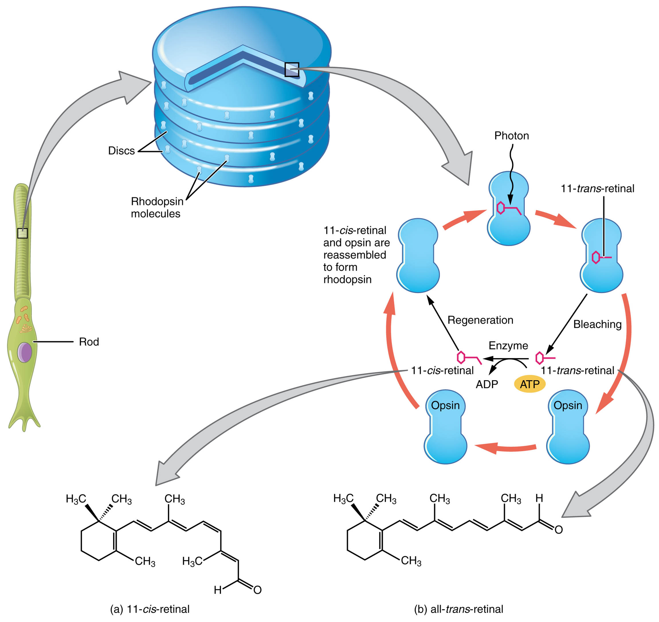
11-cis-retinal The 11-cis-retinal is the inactive isomer of retinal, present before photon interaction, bound to opsin to form rhodopsin. Its bent structure prevents signal activation until light triggers a change, initiating the visual process.
All-trans-retinal The all-trans-retinal is the active isomer formed after photoisomerization, resulting from light absorption by 11-cis-retinal. This straightened configuration triggers a conformational change in rhodopsin, sending a signal to the brain via the optic nerve.
Anatomy of Retinal Isomers
Retinal isomers are critical components of the photoreceptor cells within the retina, driving the initial step of vision. This diagram highlights the structural shift that occurs during light detection.
- The 11-cis-retinal exists in a stable, bent form, covalently linked to opsin in rod cells.
- The all-trans-retinal emerges as a linear molecule after light-induced isomerization.
- This transformation happens within the outer segments of photoreceptors, specifically in the disc membranes.
- Rhodopsin, the light-sensitive pigment, relies on these isomers for its function.
- The retina contains millions of such molecules, ensuring sensitivity across light conditions.
- The process is reversible, with enzymes converting all-trans-retinal back to 11-cis-retinal.
Physiology of Photoisomerization
Photoisomerization is the key mechanism by which retinal isomers convert light into a neural signal. This diagram illustrates the dynamic change that underpins visual transduction.
- The 11-cis-retinal absorbs a photon, causing a rapid shift to the all-trans-retinal form.
- This isomerization breaks the 11-cis double bond, altering the molecule’s shape within nanoseconds.
- The change activates rhodopsin, initiating a G-protein cascade in the photoreceptor.
- The activated state triggers the closure of sodium channels, hyperpolarizing the cell.
- This signal is then transmitted through bipolar and ganglion cells to the brain.
- The efficiency of this process allows detection of light at very low intensities.
Role of 11-cis-Retinal in Visual Readiness
The 11-cis-retinal prepares the eye for light detection with its unique structure. Its inactive state is essential for maintaining visual sensitivity.
- The bent configuration of 11-cis-retinal fits snugly into the opsin binding pocket.
- This stability prevents spontaneous activation, conserving energy until light is present.
- The molecule is regenerated in the retinal pigment epithelium after photoisomerization.
- Its presence in rods enhances low-light vision, while in cones it supports color vision.
- The synthesis involves the retinoid cycle, ensuring a constant supply.
- Deficiency in this isomer can impair dark adaptation, affecting night vision.
Role of All-trans-Retinal in Signal Activation
The all-trans-retinal drives the visual signal by altering rhodopsin’s shape post-isomerization. Its active form is crucial for translating light into perception.
- The straightened all-trans-retinal disrupts the rhodopsin complex, activating it.
- This activation releases transducin, a G-protein that amplifies the light signal.
- The molecule is subsequently released from opsin, marking the end of the active phase.
- Enzymes like retinal isomerase convert it back to 11-cis-retinal for reuse.
- The process’s speed allows rapid adaptation to changing light conditions.
- Accumulation of all-trans-retinal can lead to oxidative stress if not properly managed.
Biochemical Pathway of Retinal Isomerization
The transformation between retinal isomers involves a well-orchestrated biochemical pathway. This diagram serves as a visual guide to this critical visual process.
- Photoisomerization occurs when a photon’s energy excites an electron in 11-cis-retinal.
- The resulting all-trans-retinal triggers a conformational shift in the opsin protein.
- This shift activates the phototransduction cascade, involving cGMP and ion channels.
- The retinal pigment epithelium recycles all-trans-retinal via the visual cycle.
- Vitamin A, a precursor, is essential for maintaining retinal isomer levels.
- Disruptions in this cycle can affect the retina’s ability to regenerate 11-cis-retinal.
Clinical Relevance of Retinal Isomers
Understanding retinal isomers is vital for diagnosing and treating vision disorders. This diagram provides a foundation for exploring related conditions.
- Retinitis pigmentosa involves impaired isomerization, leading to rod cell degeneration.
- Age-related macular degeneration affects the retinoid cycle, impacting cone function.
- Excessive all-trans-retinal can contribute to retinal oxidative damage.
- Vitamin A deficiency reduces 11-cis-retinal availability, causing night blindness.
- Gene therapies aim to restore the isomerization process in genetic retinal diseases.
- Optical imaging tracks isomer levels to monitor retinal health.
- Early intervention can mitigate vision loss linked to these biochemical changes.
In conclusion, the retinal isomers depicted in this diagram are at the heart of the visual system’s ability to detect and process light. Their dynamic interplay within photoreceptors highlights the elegance of human vision, offering a rich area for exploring ocular physiology and health.

