Developmental Stages of Mandibular Molars: Radiographic Analysis of Third, Second, and First Molar Eruption
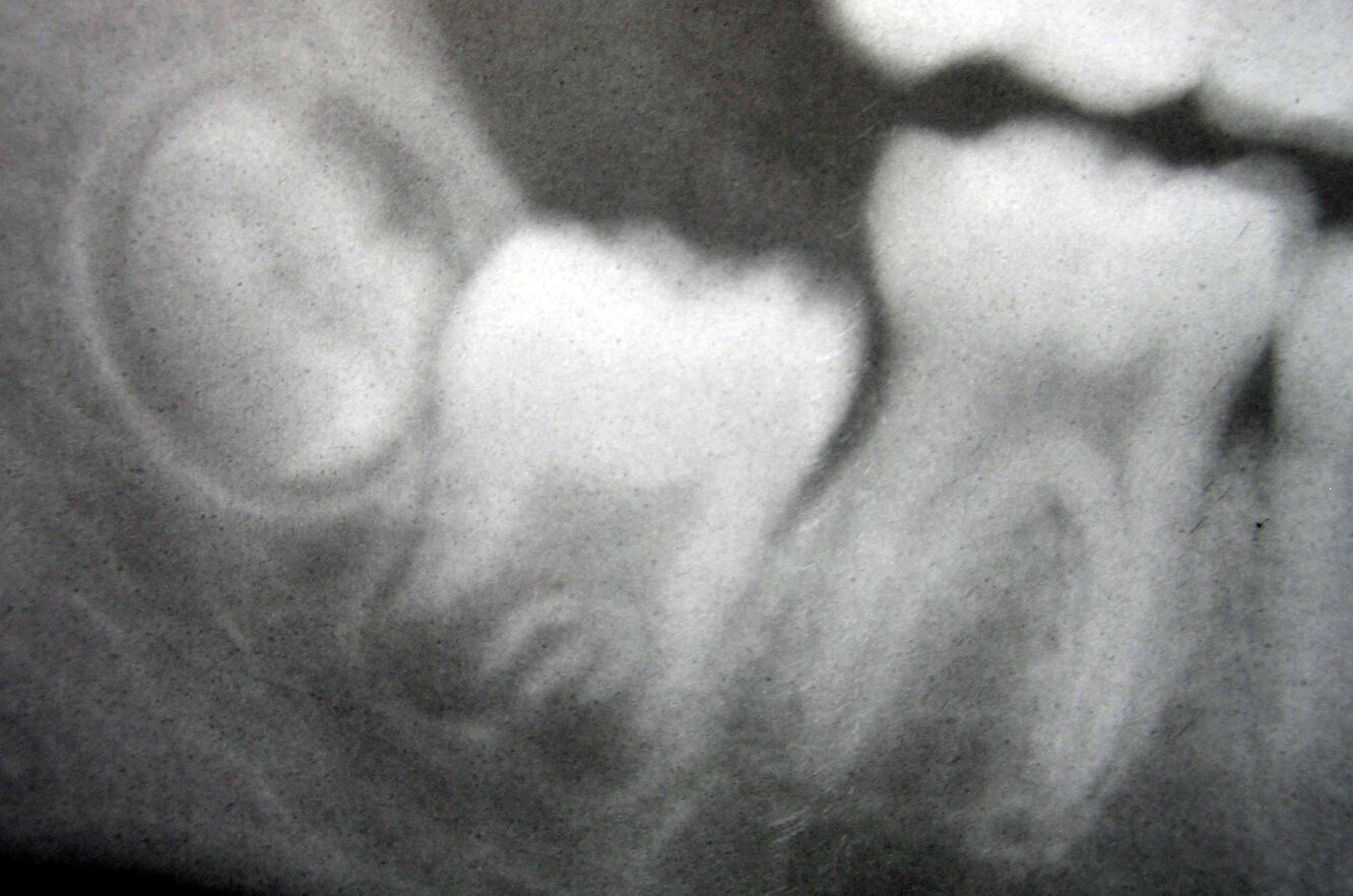
This dental radiograph provides a clear visualization of the lower right mandibular molars in varying developmental stages, captured in a periapical X-ray image. From left to right, the third molar (wisdom tooth), second molar, and first molar are visible, each representing different phases of dental development and eruption. The first molar, appearing fully erupted with complete root formation, contrasts with the second molar, which shows advanced but not complete development, while the third molar appears to be in an earlier stage of development and eruption. This radiographic presentation offers dental professionals and students valuable insights into the normal chronological development of permanent molars, their relative positions within the mandible, and the characteristic radiographic appearances that correspond to different developmental stages. Understanding these patterns is essential for age estimation, treatment planning in pediatric dentistry and orthodontics, and recognizing potential developmental anomalies that may require intervention.
Radiographic Analysis of Molar Development
The Chronology of Molar Eruption
Molar eruption follows a predictable sequence that serves as an important developmental milestone in dental practice. The timing and sequence provide crucial reference points for dental practitioners.
- First molars typically erupt around age 6-7 years, earning them the designation “6-year molars” and representing the first permanent teeth to emerge in the oral cavity.
- Second molars generally erupt around age 11-13 years, while third molars (wisdom teeth), when they do erupt, typically emerge between ages 17-25 years, though considerable variation exists.
The radiographic appearance of each molar in this image reflects these developmental timeframes. The differences in appearance demonstrate the progressive nature of tooth development.
- Root development continues for approximately 2-3 years after clinical crown eruption, which explains the variation in root formation visible among these three molars.
- The degree of root formation and apex closure provides valuable information for age estimation in forensic dentistry and pediatric patient assessment.
Radiographic Features of Developing Molars
Dental radiographs reveal distinctive features at each developmental stage that allow practitioners to assess normal progression or identify abnormalities. These features are clearly demonstrated in the image.
- The radiopaque crown structures (enamel and dentin) appear as bright white areas, while the pulp chambers and root canals present as radiolucent (darker) regions within the tooth structure.
- The surrounding periodontal ligament space appears as a thin radiolucent line around the roots, while the lamina dura (the thin layer of compact bone lining the socket) presents as a radiopaque line adjacent to this space.
Several key developmental markers are visible in the radiograph, providing information about the maturation status of each tooth. These markers help determine developmental age and potential concerns.
- The apical foramen status (open or closed) indicates the stage of root formation completion, with the first molar showing complete closure while the third molar apex remains widely open.
- The height of the alveolar bone crest around each tooth reflects the eruption status and periodontal health, showing progressive changes from the fully erupted first molar to the partially erupted third molar.
Clinical Significance of Molar Development Patterns
Diagnostic Applications in Dentistry
Radiographic evaluation of molar development provides essential information for numerous clinical applications. This type of assessment guides treatment planning across multiple dental specialties.
- In orthodontics, understanding molar development timing influences decisions regarding extraction sequences, space maintenance, and the optimal timing for treatment initiation.
- For oral surgeons, third molar development assessment is crucial for determining the appropriate time for extraction, especially when impaction is anticipated based on available space and eruption path.
Age estimation based on dental development has important applications in both clinical and forensic contexts. The predictable sequence of dental development makes it a reliable biological marker.
- Dental age assessment often proves more accurate than skeletal age estimation in children and adolescents, particularly between ages 5-17 years.
- The Demirjian method, one of the most widely used systems for dental age estimation, relies heavily on the developmental stages of molars as seen in radiographs similar to this one.
Potential Developmental Complications
The radiographic evaluation of developing molars allows for early identification of potential complications that may require intervention. Recognizing these patterns early improves treatment outcomes.
- Impaction risk assessment for third molars involves evaluating the available space, angulation, and relationship to adjacent structures such as the inferior alveolar nerve.
- Delayed eruption or abnormal development patterns may indicate systemic conditions, endocrine disorders, or localized factors that warrant further investigation.
Specific radiographic findings may signal the need for preventive or interceptive treatment. These subtle signs are important for proactive dental care.
- Mesial drift of the first molar following premature loss of primary molars can be monitored radiographically, informing decisions about space maintenance.
- Early signs of root resorption on second molars due to impacted third molars would be visible on radiographs like this one, potentially indicating the need for third molar extraction.
Radiographic Technique and Interpretation
Imaging Modalities for Molar Assessment
Various radiographic techniques are employed to evaluate molar development, each with specific advantages for different clinical scenarios. The image shown appears to be a standard periapical radiograph.
- Periapical radiographs, like the one presented, provide detailed views of individual teeth, their roots, and immediate surrounding structures, making them ideal for assessing developmental stages.
- Panoramic radiographs offer a broader view of all teeth and supporting structures, allowing for comparative assessment of development across the dentition but with less detail than periapical images.
Digital radiography has revolutionized dental imaging, offering advantages over traditional film-based techniques. Modern dental practices increasingly rely on digital systems.
- Reduced radiation exposure (typically 50-80% less than conventional film), immediate image availability, and enhancement capabilities make digital radiography particularly valuable in pediatric dentistry.
- Digital systems allow for contrast and brightness adjustment, magnification, and measurement tools that aid in precise developmental assessment.
Normal vs. Abnormal Developmental Patterns
Distinguishing between normal variations and pathological findings requires thorough knowledge of typical developmental patterns. The radiograph demonstrates normal developmental variation.
- Root morphology normally varies between individuals and tooth types, with mandibular molars typically having two roots (mesial and distal) though additional roots or fused roots may represent normal variations.
- The pulp chamber and canal size naturally decrease over time due to secondary dentin formation, with younger teeth showing larger pulp spaces as visible in the developing third molar.
Several radiographic findings would signal abnormal development requiring intervention, though these are not evident in the current image. Practitioners should remain vigilant for these signs.
- Abnormalities such as dilaceration (abrupt angular deviation of roots), taurodontism (enlarged pulp chambers with apical displacement of the pulpal floor), or congenitally missing third molars would be identifiable on radiographs.
- Pathological conditions like periapical lesions, cysts associated with developing teeth, or evidence of external root resorption would appear as radiolucent areas that disrupt the normal presentation.
Practical Applications in Dental Practice
Treatment Planning Considerations
Understanding molar developmental stages directly impacts treatment planning across multiple dental disciplines. This radiographic information guides clinical decision-making.
- In restorative dentistry, the stage of root development influences decisions regarding pulp therapy options, with immature teeth requiring different approaches such as apexogenesis or apexification rather than conventional root canal treatment.
- Orthodontic treatment timing may be optimized based on molar developmental status, with certain techniques being more effective during specific developmental windows.
The third molar presents unique management considerations due to its variable development and frequent complications. Careful monitoring through serial radiographs is often necessary.
- The decision to extract asymptomatic third molars remains controversial, with factors such as development stage, eruption path, available space, and relationship to vital structures all requiring assessment.
- Partial eruption increases the risk of pericoronitis (inflammation of the tissue surrounding the crown), with radiographic monitoring helping to identify patients at higher risk.
Educational and Communication Value
Radiographs like this one serve as valuable educational tools for both practitioners and patients. Visual representation enhances understanding and communication.
- For dental students, sequential radiographs of molar development help solidify understanding of dental embryology, eruption patterns, and age-related changes that might otherwise remain abstract concepts.
- When communicating with patients or parents of pediatric patients, these images provide visual evidence that aids in explaining developmental status and treatment recommendations.
Documentation through radiographic records provides a baseline for monitoring development over time. Sequential images allow for comparative assessment.
- Serial radiographs taken at appropriate intervals enable tracking of developmental progress and early identification of deviations from expected patterns.
- Digital record systems facilitate longitudinal comparison, allowing practitioners to overlay images from different timepoints to assess subtle changes in position or development.
Conclusion
This radiographic image of mandibular molars at different developmental stages serves as an excellent reference for understanding the natural progression of dental development. The visualization of the third, second, and first molars side by side demonstrates the sequential nature of tooth formation and eruption that dental practitioners rely upon for numerous clinical assessments. From the fully developed first molar to the partially formed third molar, each tooth tells part of the developmental story, providing insights into approximate dental age and potential future concerns. For dental professionals, such radiographs are invaluable tools for patient assessment, treatment planning, and monitoring developmental milestones. They also serve as excellent educational resources for dental students learning to recognize the radiographic features associated with different developmental stages. Understanding these patterns enables more accurate diagnosis, improved treatment timing, and better recognition of developmental anomalies that may require intervention.
- Radiographic Analysis of Mandibular Molar Development: Interpreting First, Second, and Third Molar Eruption Stages
- Developmental Chronology of Lower Molars: A Radiographic Guide for Dental Professionals
- Understanding Molar Eruption Patterns: Radiographic Evaluation of Mandibular First, Second, and Third Molars
- Comparative Radiographic Assessment of Permanent Molar Development in the Mandibular Arch
- Dental Age Estimation Through Radiographic Analysis of Mandibular Molar Developmental Stages

