The pupillary reflex pathways diagram provides a detailed look at how the eyes adapt to varying light conditions through intricate neural circuits. This chart highlights the interplay between the sympathetic and parasympathetic systems, which respectively dilate or constrict the pupil to optimize vision and protect the retina. Exploring these pathways offers a deeper understanding of ocular reflexes and their significance in maintaining visual health.
Labeled Components in the Diagram
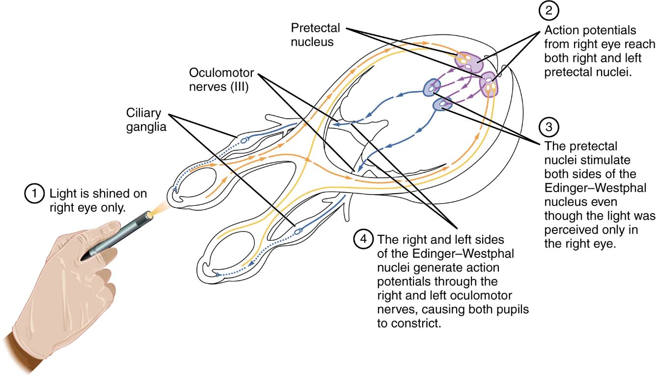
Light is shined on right eye only The light is shined on right eye only depicts the initial stimulus where light activates the retina of the right eye. This triggers a reflex response that involves both eyes due to bilateral neural connections.
Pretectal nucleus The pretectal nucleus is a midbrain structure that receives light signals from the retina via the optic nerve. It processes this input and relays it to initiate the pupillary reflex on both sides.
Oculomotor nerves (III) The oculomotor nerves (III) are cranial nerves that carry parasympathetic signals from the Edinger-Westphal nucleus to the ciliary ganglia. They play a key role in controlling pupil constriction.
Ciliary ganglia The ciliary ganglia are peripheral ganglia where preganglionic parasympathetic neurons synapse with postganglionic neurons. These ganglia send signals to the iris muscles to adjust pupil size.
Action potentials from right eye reach both right and left pretectal nuclei The action potentials from right eye reach both right and left pretectal nuclei indicate bilateral signal transmission from the stimulated eye. This ensures a coordinated response affecting both pupils.
The pretectal nuclei stimulate both sides of the Edinger-Westphal nucleus even though the light was perceived only in the right eye The the pretectal nuclei stimulate both sides of the Edinger-Westphal nucleus even though the light was perceived only in the right eye reflects the consensual reflex. This bilateral activation ensures both pupils constrict simultaneously.
The right and left sides of the Edinger-Westphal nuclei generate action potentials through the right and left oculomotor nerves, causing both pupils to constrict The the right and left sides of the Edinger-Westphal nuclei generate action potentials through the right and left oculomotor nerves, causing both pupils to constrict details the final step. This action results in pupil constriction to protect the retina from excessive light.
Anatomy of the Pupillary Reflex
The pupillary reflex is a protective mechanism that adjusts pupil size based on light intensity. This reflex involves a coordinated effort between the eyes and the brain’s neural network.
- The retina contains photoreceptors that detect light and send signals through ganglion cells.
- Optic nerve fibers carry these signals to the pretectal nucleus in the midbrain.
- The Edinger-Westphal nucleus, a parasympathetic center, integrates these signals.
- Oculomotor nerves (III) transmit signals to ciliary ganglia on both sides.
- Postganglionic neurons from the ganglia innervate the iris’s circular muscles.
- The consensual reflex ensures both pupils respond, even if only one eye is stimulated.
Physiological Mechanisms of the Reflex
The physiological response to light involves rapid neural signaling and muscle contraction. This process maintains optimal light levels for vision while safeguarding the retina.
- Light activates retinal ganglion cells, generating action potentials.
- These potentials travel via the optic nerve to the pretectal nucleus.
- The pretectal nucleus sends excitatory signals to both Edinger-Westphal nuclei.
- Preganglionic parasympathetic neurons release acetylcholine at the ciliary ganglia.
- Postganglionic neurons stimulate circular iris muscles to contract, constricting the pupil.
- The reflex completes within milliseconds, demonstrating efficient neural coordination.
Role of the Pretectal and Edinger-Westphal Nuclei
The pretectal and Edinger-Westphal nuclei are central to the pupillary reflex’s integration. Their roles ensure a swift and bilateral response to light changes.
- The pretectal nucleus acts as an intermediary, processing retinal input.
- It projects to both Edinger-Westphal nuclei, enabling the consensual reflex.
- The Edinger-Westphal nucleus contains preganglionic parasympathetic neurons.
- These neurons generate action potentials in response to bright light.
- Bilateral activation prevents unequal pupil responses, maintaining symmetry.
- Damage to these nuclei can impair the reflex, indicating neurological issues.
Clinical Significance of the Pupillary Reflex
The pupillary reflex serves as a diagnostic tool for assessing neurological and ocular health. Its evaluation can reveal underlying conditions affecting the nervous system.
- A sluggish reflex may suggest optic nerve damage or midbrain lesions.
- Unequal pupil responses can indicate cranial nerve III palsy.
- The absence of constriction might point to severe brain injury or coma.
- The swinging flashlight test checks for relative afferent pupillary defect.
- Adie’s syndrome, involving a tonic pupil, affects reflex speed.
- Regular reflex testing is vital in trauma and neurological assessments.
Comparison with Sympathetic Control
While this diagram focuses on parasympathetic control, sympathetic input also influences pupil size. Understanding this balance enhances comprehension of ocular regulation.
- Sympathetic activation dilates the pupil via radial muscle contraction.
- The superior cervical ganglion mediates this response in dim light.
- Norepinephrine release stimulates dilation, contrasting with acetylcholine’s effect.
- The parasympathetic system dominates in bright light for constriction.
- Both systems maintain a dynamic equilibrium for visual adaptation.
- Imbalances can lead to conditions like Horner’s syndrome or anisocoria.
Advances in Reflex Research
Recent advancements in neurophysiology offer new insights into the pupillary reflex. These developments pave the way for improved diagnostics and treatments.
- High-resolution imaging tracks neural activity in the pretectal nucleus.
- Optogenetics manipulates reflex pathways in experimental models.
- Neuroprotective agents aim to preserve ganglion cell function.
- Artificial intelligence analyzes reflex patterns for early detection.
- Surgical interventions target oculomotor nerve damage.
- Wearable devices monitor pupil responses in real-time.
The pupillary reflex pathways demonstrate the body’s remarkable ability to protect and adapt its vision through neural precision. By studying these mechanisms, professionals can enhance diagnostic accuracy and develop targeted therapies, ultimately improving patient outcomes and visual health.

