Direct activation through ionotropic receptors is a cornerstone of rapid neural communication, enabling immediate responses to stimuli. This article explores the process depicted in the provided diagram, shedding light on how neurotransmitters trigger ion channels to alter membrane potential. By understanding this mechanism, one can appreciate the efficiency and precision of neuronal signaling in the nervous system.
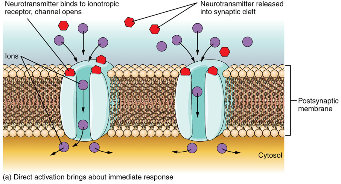
Neurotransmitter binds to ionotropic receptor, channel opens: This label illustrates the binding of a neurotransmitter to an ionotropic receptor, which promptly opens an ion channel. The rapid change allows ions to flow, altering the membrane potential almost instantly to facilitate signal transmission.
Neurotransmitter released into synaptic cleft: This indicates the release of neurotransmitters from the presynaptic neuron into the synaptic cleft. These chemical messengers travel across this gap to interact with receptors on the postsynaptic membrane, initiating the response.
Ions: This label highlights the movement of ions, such as sodium or chloride, through the opened ion channels. The direction and type of ion flow determine whether the postsynaptic neuron is excited or inhibited.
Postsynaptic membrane: This is the receiving surface of the neuron, embedded with ionotropic receptors that respond to neurotransmitters. Its activation leads to immediate changes in the neuron’s electrical state.
Cytosol: This refers to the intracellular fluid where ions enter or exit upon channel opening. It serves as the medium where the immediate effects of ion movement are felt within the neuron.
Anatomy of Direct Activation
The structure involved in direct activation is designed for swift signal transmission. Each component plays a specific role in ensuring rapid neuronal responses.
- The postsynaptic membrane contains ionotropic receptors that are key to this process.
- The synaptic cleft acts as the pathway for neurotransmitter diffusion.
- Ions move through channels to influence the neuron’s internal environment.
- The cytosol is where these ionic changes impact cellular function.
- This anatomical setup supports the speed required for reflex actions.
Physiological Process of Ionotropic Activation
The physiological mechanism of ionotropic receptors ensures a fast and direct response. This process is critical for the nervous system’s ability to react promptly to stimuli.
- Neurotransmitter binding to an ionotropic receptor opens the channel immediately.
- Ions, such as sodium, rush into the cytosol, depolarizing the postsynaptic membrane.
- This depolarization can trigger an action potential if the threshold is reached.
- The response is short-lived, ceasing once the neurotransmitter is cleared.
- Speed is enhanced by the direct nature of this ion channel activation.
Clinical Relevance and Neural Function
While direct activation itself is not a disease, its dysfunction can contribute to neurological issues. Proper functioning of ionotropic receptors is vital for healthy neural communication.
- Malfunctions in ionotropic receptors are associated with conditions like epilepsy.
- Imbalances in ion flow can lead to hyperexcitability or paralysis.
- The postsynaptic membrane’s receptor density affects disease susceptibility.
- Drugs targeting these receptors can treat disorders by modulating neurotransmitter activity.
- Understanding this process aids in developing therapies for neural dysregulation.
Direct activation via ionotropic receptors is a fascinating aspect of neural physiology, enabling the nervous system to respond swiftly to environmental changes. The diagram clearly depicts how neurotransmitter release and ion movement work together to produce an immediate effect, offering a valuable tool for learning. By mastering these concepts, one can gain a deeper understanding of the intricate balance required for effective neural signaling and its broader impact on bodily functions.

