The human eye is a remarkable organ, intricately designed with distinct chambers and layers that support vision. This lateral view highlights the eye’s anatomical structure, including its muscular, vascular, and neural components, offering a comprehensive look at its functionality.
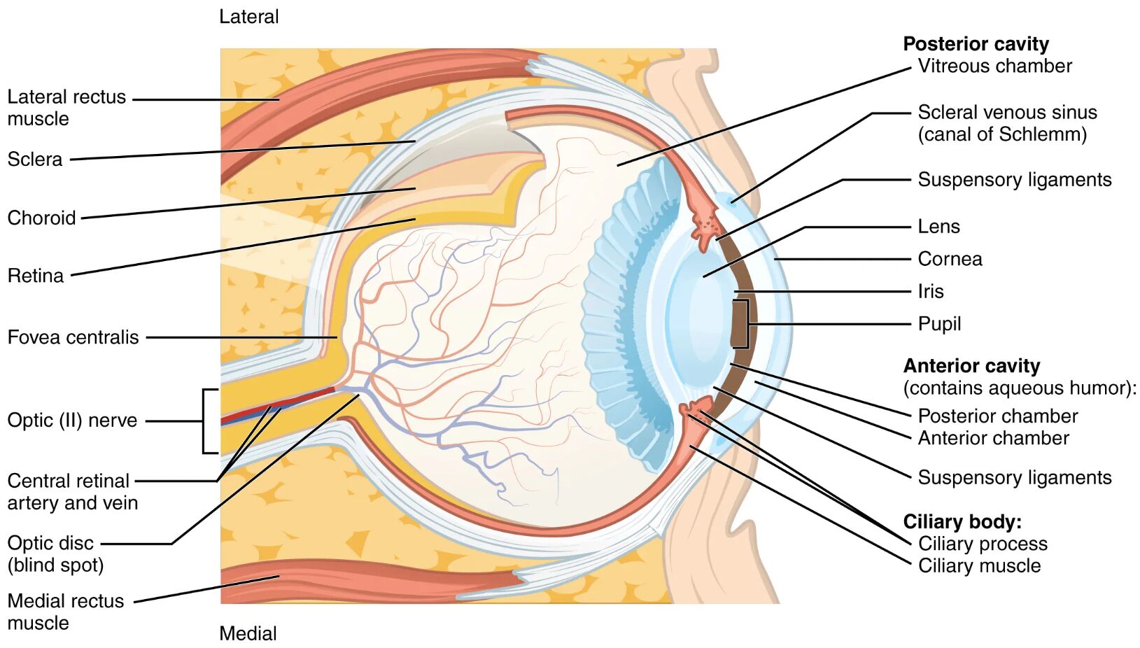
Lateral rectus muscle The lateral rectus muscle abducts the eye, moving it outward away from the nose. Controlled by the abducens nerve, it plays a critical role in lateral eye movements.
Sclera The sclera is the tough, white outer layer of the eyeball, providing structural support and protection. It transitions into the cornea at the front, maintaining the eye’s shape.
Choroid The choroid is a vascular layer beneath the sclera, rich in blood vessels that nourish the retina. It also contains pigment to reduce light scatter within the eye.
Retina The retina is the innermost neural layer, containing photoreceptors that convert light into electrical signals. It features three cell layers and two synaptic layers, with the fovea as a key visual center.
Fovea centralis The fovea centralis is a small pit in the retina’s center, packed with cones for sharp central vision. It is the point of clearest visual acuity, ideal for detailed tasks.
Optic (II) nerve The optic (II) nerve transmits visual information from the retina to the brain. It exits the eye at the optic disc, forming the connection for visual processing.
Central retinal artery and vein The central retinal artery and vein supply blood to and drain from the retina, ensuring its metabolic needs. They enter and exit through the optic disc, supporting retinal function.
Optic disc (blind spot) The optic disc (blind spot) is the area where the optic nerve exits, lacking photoreceptors. This creates a natural blind spot in each eye’s visual field.
Medial rectus muscle The medial rectus muscle adducts the eye, moving it inward toward the nose. Innervated by the oculomotor nerve, it balances lateral movements for alignment.
Vitreous chamber The vitreous chamber is the large posterior cavity filled with vitreous humor, a gel-like substance. It maintains eye shape and supports the retina by providing pressure.
Scleral venous sinus (canal of Schlemm) The scleral venous sinus (canal of Schlemm) drains aqueous humor from the anterior chamber, regulating intraocular pressure. It is located at the junction of the sclera and cornea.
Suspensory ligaments The suspensory ligaments hold the lens in place, connecting it to the ciliary body. They adjust lens shape for focusing on near or distant objects.
Lens The lens is a transparent structure behind the iris that refracts light onto the retina. Its elasticity, controlled by the ciliary muscle, allows for accommodation.
Cornea The cornea is the clear front layer of the eye, refracting light to focus it on the retina. It is avascular, relying on tears and aqueous humor for nourishment.
Iris The iris is the colored part of the eye, controlling pupil size to regulate light entry. It contains muscles that respond to light intensity changes.
Pupil The pupil is the opening in the iris through which light enters the eye. Its diameter adjusts to optimize visual clarity in varying light conditions.
Anterior chamber (contains aqueous humor) The anterior chamber (contains aqueous humor) is the front cavity between the cornea and iris, filled with aqueous humor. This fluid nourishes the cornea and lens while maintaining pressure.
Posterior chamber The posterior chamber is the small space behind the iris and in front of the lens, also filled with aqueous humor. It facilitates fluid circulation within the eye.
Ciliary body: Ciliary process The ciliary body: Ciliary process secretes aqueous humor and supports the lens via suspensory ligaments. These processes are part of the ciliary body’s role in fluid production.
Ciliary muscle The ciliary muscle adjusts the lens shape by contracting or relaxing, enabling accommodation for near or far vision. It is controlled by the parasympathetic nervous system.
Structure of the Eye’s Layers
The eye’s wall comprises three main layers, each with a distinct role in vision and protection. This lateral view reveals how these layers integrate with internal structures to support sight.
- The fibrous tunic, including the sclera and cornea, forms the outer protective layer.
- The vascular tunic, or choroid, supplies nutrients and regulates light within the eye.
- The neural tunic, or retina, contains photoreceptors and neural cells for light processing.
- The fovea centralis within the retina enhances central vision with a high density of cones.
- The optic (II) nerve and central retinal artery and vein connect the retina to the brain and blood supply.
- The optic disc (blind spot) marks the nerve exit, a natural gap in the visual field.
- Supporting tissues like the medial rectus muscle and lateral rectus muscle stabilize the eyeball.
Functions of Eye Chambers and Fluids
The eye’s chambers and fluids maintain its shape and support optical functions. This lateral perspective highlights their critical roles in vision.
- The vitreous chamber holds vitreous humor, providing structural support and a clear medium for light.
- The anterior chamber (contains aqueous humor) nourishes the cornea and maintains intraocular pressure.
- The posterior chamber circulates aqueous humor, aiding in pressure regulation.
- The scleral venous sinus (canal of Schlemm) drains excess aqueous humor to prevent glaucoma.
- The lens, supported by suspensory ligaments, focuses light with adjustable curvature.
- The ciliary body: Ciliary process produces aqueous humor, while the ciliary muscle adjusts focus.
- These components work together to ensure a stable and clear visual field.
Role of the Retina and Fovea
The retina serves as the eye’s light-sensitive layer, with the fovea centralis as its focal point. Their structure supports high-acuity vision critical for detailed tasks.
- The retina contains rods for low-light vision and cones for color and detail, concentrated in the fovea.
- The fovea centralis lacks blood vessels, maximizing light access to photoreceptors.
- Three cell layers—photoreceptors, bipolar cells, and ganglion cells—process visual signals.
- The optic (II) nerve carries these signals, exiting at the optic disc (blind spot).
- The central retinal artery and vein ensure oxygen and nutrient delivery to the retina.
- Damage to the fovea can lead to central vision loss, though this image shows normal anatomy.
Muscular and Vascular Support
Muscles and vascular structures stabilize and nourish the eye, as seen in this lateral view. Their coordination is vital for eye movement and health.
- The lateral rectus muscle and medial rectus muscle control horizontal eye alignment.
- The choroid provides a rich blood supply, preventing retinal detachment risks.
- The sclera offers mechanical support, anchoring muscle insertions.
- The ciliary muscle adjusts the lens for accommodation, aided by suspensory ligaments.
- The scleral venous sinus (canal of Schlemm) regulates fluid outflow to maintain pressure.
- Inflammation or injury to these structures can affect vision, but this diagram depicts healthy conditions.
Clinical Relevance of Eye Anatomy
Understanding the eye’s lateral anatomy assists in diagnosing and managing ocular conditions. This image provides a baseline for assessing normal structure and function.
- Elevated intraocular pressure, linked to scleral venous sinus (canal of Schlemm) dysfunction, can lead to glaucoma.
- Retinal detachment may involve the retina and choroid, requiring surgical intervention.
- The fovea centralis damage can cause macular degeneration, affecting central vision.
- Muscle imbalances, such as with the lateral rectus muscle, may result in strabismus.
- The optic (II) nerve issues can lead to optic neuritis, impacting visual fields.
- Regular eye exams monitor these structures for early detection of abnormalities.
- Treatments like laser therapy or surgery address specific anatomical concerns.
In conclusion, the eye’s lateral view reveals a complex interplay of chambers, layers, and muscles that sustain vision. This anatomical insight underscores the eye’s remarkable ability to adapt and function, making it a fascinating subject for exploring human physiology.

