Delve into the fundamental architecture of the human brain by exploring the distinct roles of gray matter and white matter, as revealed in a cadaveric brain section. This article explains how gray matter forms the brain’s outer cortex, responsible for processing information, while white matter facilitates rapid communication across different brain regions. Gain crucial insights into these critical components for a deeper understanding of neurological function and health.
Gray matter: This component primarily consists of neuronal cell bodies, unmyelinated axons, dendrites, and all nerve synapses. It forms the outer cortex of the cerebrum and cerebellum, as well as the deeper nuclei within the brain, and is largely responsible for processing information, perception, memory, and voluntary movement.
White matter: Composed mainly of myelinated axons, white matter acts as the communication superhighway of the brain, connecting various gray matter areas to each other and to other parts of the body. The myelin sheath, a fatty substance, insulates these axons, enabling the rapid and efficient transmission of electrical signals.
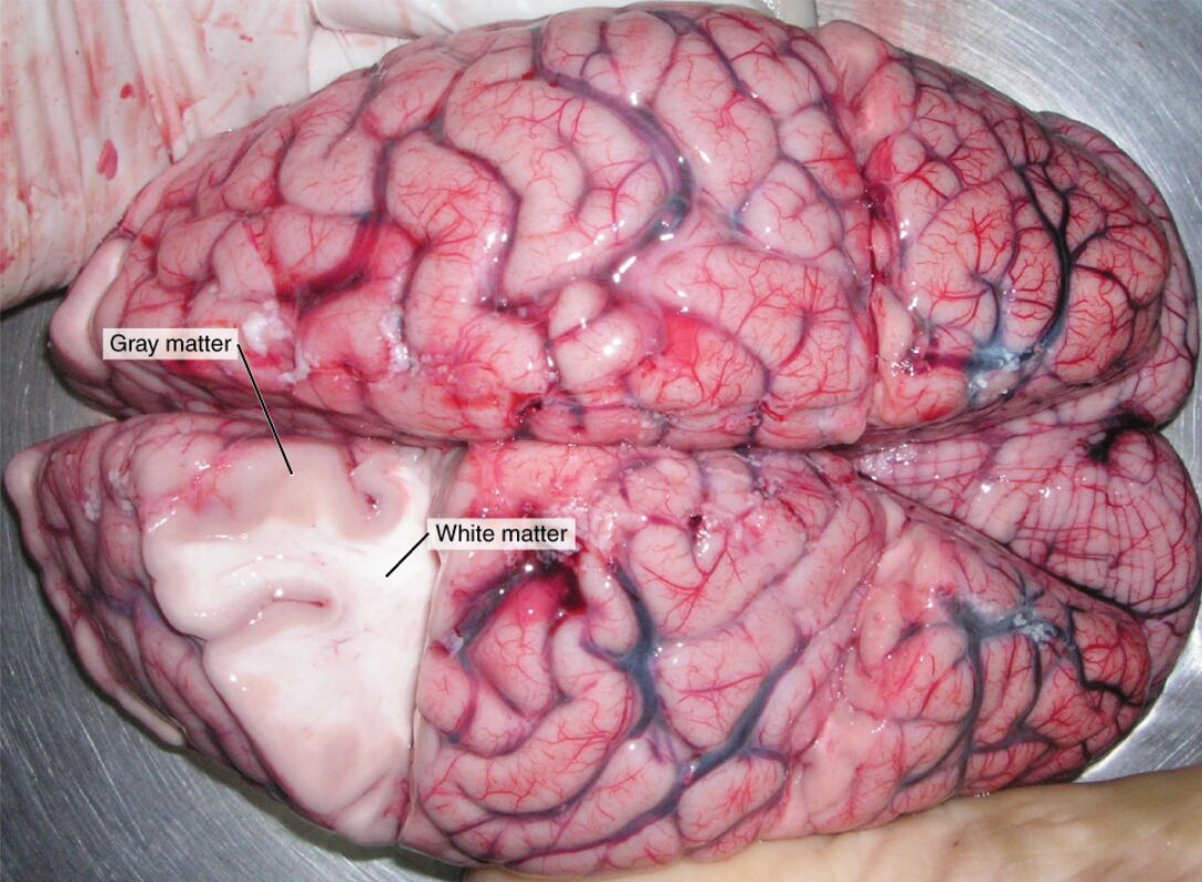
The human brain is an extraordinarily complex organ, serving as the command center for the entire nervous system. Its intricate structure and functional organization are what allow for consciousness, thought, emotion, and control over virtually all bodily functions. At a fundamental level, the brain can be broadly categorized into two main tissue types: gray matter and white matter. These distinct components, though working in concert, have specialized roles crucial for information processing and transmission. The image, showing a cadaveric brain with a section removed, provides a stark visual representation of this critical anatomical distinction, revealing the darker, convoluted surface of the gray matter overlying the lighter-toned white matter.
The Role of Gray Matter
Gray matter is found on the surface of the cerebrum and cerebellum, forming the cerebral cortex, and also in deeper brain structures like the thalamus, hypothalamus, and basal ganglia. It is characterized by its high concentration of neuronal cell bodies, dendrites, unmyelinated axons, glial cells, and capillaries. This dense collection of nerve cell bodies is where most of the brain’s computational work occurs. Functions such as conscious thought, memory, learning, attention, language processing, and sensory perception are primarily attributed to the intricate neural networks within the gray matter. Its convoluted surface, with gyri (ridges) and sulci (grooves), significantly increases the surface area, allowing for a vast number of neurons.
The Significance of White Matter
In contrast to gray matter, white matter is predominantly composed of myelinated axons. Myelin, a fatty substance produced by oligodendrocytes in the central nervous system, acts as an electrical insulator around the axons. This insulation is crucial for increasing the speed and efficiency of electrical signal transmission between different areas of gray matter and between the brain and other parts of the body. White matter pathways can be categorized into association fibers (connecting different parts of the same hemisphere), commissural fibers (connecting the two hemispheres, like the corpus callosum), and projection fibers (connecting the cerebrum to lower brain areas and the spinal cord). These extensive networks facilitate rapid communication and coordination across the brain.
Interdependence and Neurological Health
The dynamic interplay between gray and white matter is essential for all neurological functions. Gray matter processes information, while white matter ensures that this information is efficiently relayed to the appropriate regions for further processing or action. Damage to either type of matter can have profound neurological consequences. For instance, neurodegenerative diseases like Alzheimer’s often involve the loss of gray matter, leading to cognitive decline. Conditions affecting myelin, such as multiple sclerosis, impair white matter’s ability to transmit signals, resulting in a range of motor, sensory, and cognitive deficits. Maintaining the health and integrity of both gray and white matter is therefore paramount for optimal brain function.
In conclusion, the distinct yet interconnected roles of gray matter and white matter form the bedrock of the brain’s complex functionality. Gray matter, with its neuronal cell bodies, serves as the primary processing center, while white matter’s myelinated axons act as rapid communication pathways. A comprehensive understanding of these fundamental components is indispensable for grasping the intricacies of neurological processes, from basic sensory perception to higher-level cognitive functions, and for addressing the challenges posed by various neurological disorders.

