The synapse serves as the vital connection point between a neuron and its target cell, facilitating the transmission of signals across the nervous system. This article explores the intricate structure and function of the synapse as depicted in the provided diagram, offering a comprehensive look at how neurotransmitters bridge the gap between neurons. By delving into this process, one can better grasp the foundation of neural communication and its broader implications.
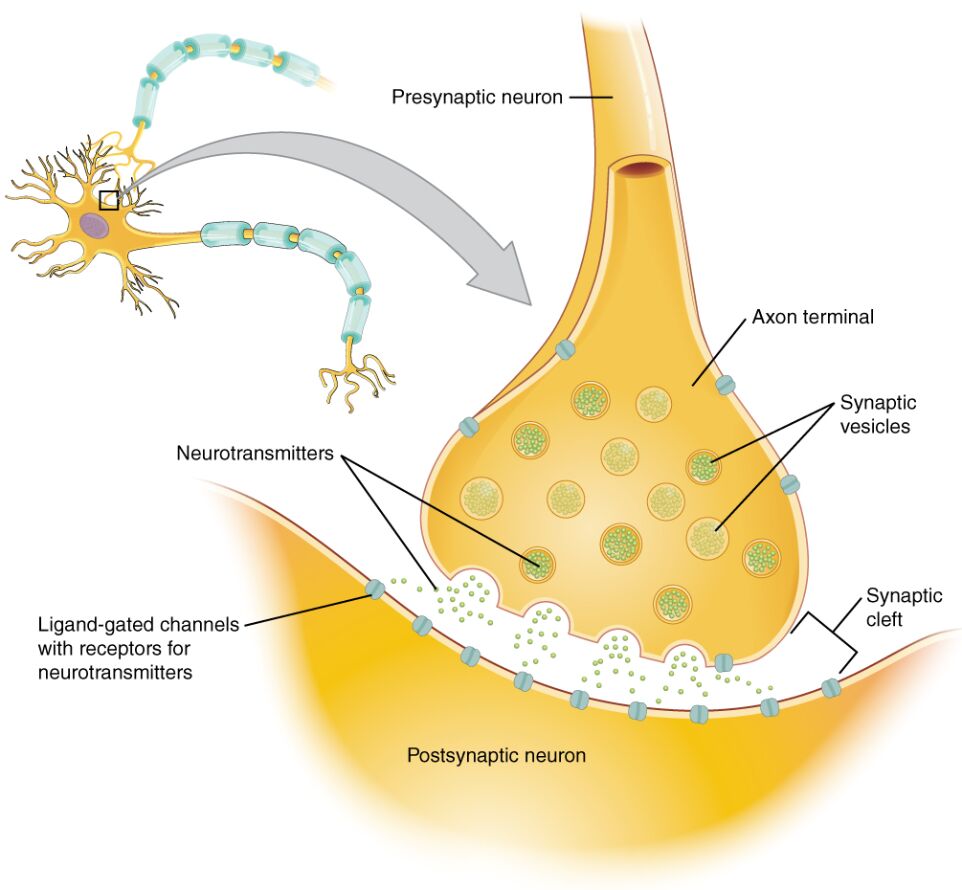
Presynaptic neuron: This is the neuron that sends the signal, with its axon extending to the synapse. It releases neurotransmitters into the synaptic cleft to communicate with the postsynaptic neuron.
Postsynaptic neuron: This is the receiving neuron or target cell, equipped with receptors to detect neurotransmitters released from the presynaptic neuron. The response of this neuron depends on the type and amount of neurotransmitter binding.
Axon terminal: This is the specialized end of the presynaptic neuron’s axon, where synaptic vesicles are stored and neurotransmitter release is initiated. It plays a crucial role in converting electrical signals into chemical signals.
Synaptic vesicles: These small sacs within the axon terminal contain neurotransmitters, ready to be released upon calcium ion entry. Their fusion with the membrane allows the release of neurotransmitters into the synaptic cleft.
Neurotransmitters: These chemical messengers are released from the presynaptic neuron and diffuse across the synaptic cleft to bind to receptors on the postsynaptic neuron. Their action can be excitatory or inhibitory, influencing the next step in signal transmission.
Ligand-gated channels with receptors for neurotransmitters: These channels on the postsynaptic neuron open when neurotransmitters bind, allowing ions like sodium or chloride to flow. This ion movement alters the membrane potential, triggering a response in the postsynaptic neuron.
Synaptic cleft: This narrow gap separates the presynaptic and postsynaptic neurons, through which neurotransmitters travel. It is a critical space where the chemical signal is transmitted and subsequently cleared.
Anatomy of the Synapse
The synapse’s structure is elegantly designed to ensure efficient signal transmission. Each component works in harmony to facilitate communication between neurons.
- The presynaptic neuron contains the machinery to synthesize and store neurotransmitters.
- The axon terminal houses synaptic vesicles, which are essential for neurotransmitter release.
- The synaptic cleft acts as a conduit, allowing rapid diffusion of neurotransmitters.
- The postsynaptic neuron features receptors that detect and respond to these chemicals.
- Ligand-gated channels regulate ion flow, influencing the electrical state of the neuron.
Physiological Process of Synaptic Transmission
Synaptic transmission involves a series of precise steps that convert electrical signals into chemical signals. This process is fundamental to understanding how neurons communicate.
- An action potential reaches the axon terminal, triggering calcium ion influx.
- Calcium entry causes synaptic vesicles to fuse with the membrane, releasing neurotransmitters.
- Neurotransmitters diffuse across the synaptic cleft to bind to ligand-gated channels.
- This binding opens channels, allowing ion movement that can depolarize or hyperpolarize the postsynaptic neuron.
- Clearance mechanisms, such as enzymatic degradation, ensure the synapse returns to its resting state.
Clinical Relevance and Neural Function
While the synapse itself is not a disease entity, its dysfunction can lead to various neurological conditions. Proper synaptic function is essential for maintaining neural health and coordination.
- Disruptions in neurotransmitter release are linked to disorders like Parkinson’s disease.
- Imbalances in ligand-gated channels can contribute to epilepsy or anxiety disorders.
- The synaptic cleft’s integrity affects signal speed and accuracy in neural pathways.
- Therapeutic drugs often target synaptic vesicles or receptors to modulate transmission.
- Understanding this process aids in developing treatments for neurodegenerative conditions.
The synapse is a remarkable junction that underpins the complexity of the nervous system, enabling everything from basic reflexes to higher cognitive functions. The diagram provides a clear visual of how presynaptic neuron, postsynaptic neuron, and associated structures work together, offering a solid foundation for exploring neural signaling. By mastering these concepts, one can appreciate the delicate balance required for effective communication and the potential impact of its disruption on overall health.

