Understanding the Skeletal Framework of the Human Nose: An In-Depth Anatomical Overview
The human nose serves as a vital gateway for respiration, olfaction, and even aesthetic facial harmony, with its skeletal structure providing both support and flexibility. Composed of a blend of bones and cartilages, this intricate framework ensures the nose can withstand daily stresses while adapting to various functions like filtering air and enhancing vocal resonance. Exploring the anatomy through diagrams reveals how these components interconnect, offering insights into both form and function that are essential for appreciating overall facial physiology.
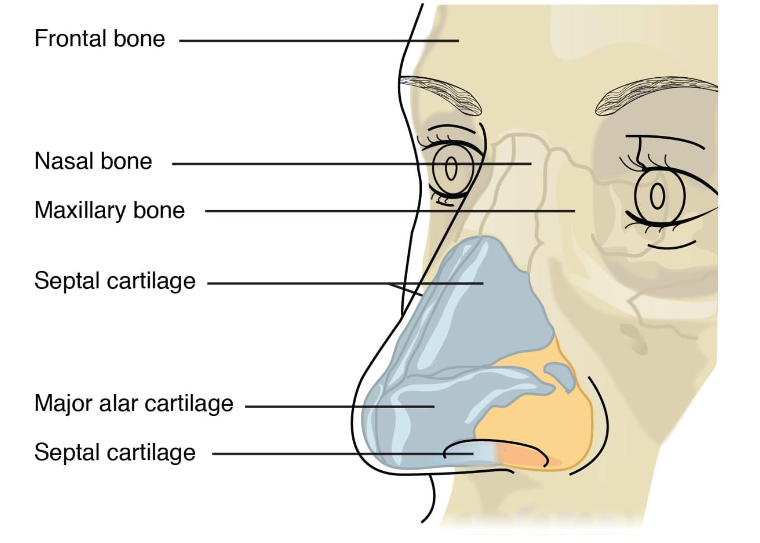
Key Anatomical Labels in the Diagram
This section breaks down each labeled element from the provided diagram, highlighting their positions and roles. Understanding these parts individually helps in grasping the nose’s overall architecture.
Frontal bone: The frontal bone forms the upper boundary of the nasal region, connecting seamlessly with the nasal bones below. It contributes to the forehead and the roof of the orbits, providing structural integrity to protect the underlying brain and eyes from trauma.
Nasal bone: Positioned at the bridge of the nose, the nasal bones are paired structures that create the bony prominence often referred to as the nasal dorsum. They articulate with the frontal bone superiorly and the maxillary bones laterally, playing a key role in defining the nose’s external shape and supporting the nasal cavity’s entrance.
Maxillary bone: The maxillary bone, also known as the maxilla, forms the lateral walls and part of the floor of the nasal cavity, extending to support the upper teeth. It interfaces with the nasal bones and contributes to the nasal aperture, ensuring stability for breathing and facial expressions.
Septal cartilage: Located centrally within the nose, the septal cartilage divides the nasal cavity into two symmetrical nostrils, forming the anterior portion of the nasal septum. It provides flexible support that allows for slight movement during breathing, while also aiding in directing airflow efficiently through the nasal passages.
Major alar cartilage: The major alar cartilage shapes the tip and nostrils of the nose, consisting of paired structures that form the lateral and medial crura. This cartilage is crucial for maintaining the nostril’s patency, preventing collapse during inhalation and contributing to the nose’s aesthetic contour.
Sesamoid cartilage: Small and variable in number, sesamoid cartilages are embedded within the soft tissues lateral to the major alar cartilage. They enhance the flexibility of the nasal ala, assisting in subtle adjustments during facial movements and respiration.
The Bony Components of Nasal Anatomy
The bony elements of the nose provide a rigid foundation that integrates with the broader facial skeleton. These structures not only support the soft tissues but also protect internal airways from external impacts.
- The frontal bone extends downward to meet the nasal bones, creating a seamless transition from the forehead to the nose.
- Paired nasal bones vary in size among individuals, influencing ethnic differences in nasal shapes.
- The maxillary bone houses the maxillary sinuses, which lighten the skull and humidify inhaled air.
- Fractures to these bones, often from trauma, can lead to deviations in the nasal septum.
- Surgical interventions like rhinoplasty frequently address bony irregularities for functional or cosmetic reasons.
Cartilaginous Structures and Their Functions
Cartilages in the nose offer elasticity, allowing the structure to adapt without breaking under pressure. Unlike bones, they do not ossify with age, preserving lifelong flexibility.
- The septal cartilage, primarily hyaline in type, receives blood supply from branches of the facial artery.
- Major alar cartilages form a tripod-like support at the nasal tip, essential for aesthetic balance.
- Sesamoid cartilages, though small, prevent pinching of the nostrils during forceful exhalation.
- These cartilages are prone to deviation in conditions like septal hematoma, affecting airflow.
- Growth of nasal cartilages continues into adulthood, sometimes leading to changes in nasal appearance over time.
Physiological Importance of the Nasal Skeleton
The nasal skeleton plays a multifaceted role in human physiology beyond mere structure. It interacts with mucous membranes to filter, warm, and humidify air before it reaches the lungs.
- Proper alignment of the nasal bones and cartilages ensures efficient turbulent airflow, trapping particles.
- The septal cartilage’s position influences olfactory nerve function, impacting smell perception.
- Maxillary bone integration with the palate supports phonation, aiding clear speech production.
- In evolutionary terms, the human nose’s prominence enhances sensory capabilities compared to other primates.
- Disruptions in this framework, such as congenital anomalies, can lead to breathing difficulties requiring medical attention.
Clinical Relevance and Common Issues
While the diagram depicts a healthy nasal structure, real-world applications often involve addressing deviations or injuries. Knowledge of these components aids in diagnosing and treating nasal disorders effectively.
- Nasal bone fractures are common in sports injuries, potentially causing cosmetic deformities.
- Septal cartilage deviations can result from birth trauma or accidents, leading to chronic congestion.
- Alar cartilage weakness may contribute to nasal valve collapse, a frequent complaint in aging populations.
- Reconstructive procedures rely on understanding sesamoid positions to restore natural contours.
- Preventive measures, like wearing protective gear, help maintain the integrity of these delicate structures.
Integration with Surrounding Facial Anatomy
The nose does not exist in isolation but connects intricately with adjacent facial features. This interconnectedness ensures coordinated functions across the craniofacial region.
- The frontal bone links to the supraorbital ridge, protecting the eyes while framing the nose.
- Maxillary bones articulate with zygomatic bones, forming the cheek’s prominence.
- Cartilaginous elements blend with skin and muscles, enabling expressions like flaring nostrils.
- Blood supply from the external carotid artery nourishes these tissues, promoting healing.
- Nerve innervation via the trigeminal nerve provides sensation, crucial for reflex actions like sneezing.
In summary, the skeletal features of the human nose represent a remarkable balance of strength and adaptability, essential for daily life and overall health. By delving into its components through diagrams like this, one gains a deeper appreciation for how this central facial feature contributes to both function and form, underscoring the elegance of human anatomy.

