The posterior pituitary plays a crucial role in the endocrine system, acting as a storage and release site for vital hormones produced in the hypothalamus. This diagram illustrates the intricate connection between the brain and the pituitary gland, highlighting the pathways of oxytocin (OT) and antidiuretic hormone (ADH) as they travel and function within the body. Exploring this structure offers valuable insights into hormonal regulation and its impact on overall health.
Labels Introduction
- Brain The brain serves as the central command hub, housing the hypothalamus which initiates hormone production. Its complex network of neurons coordinates various bodily functions, including those managed by the posterior pituitary.
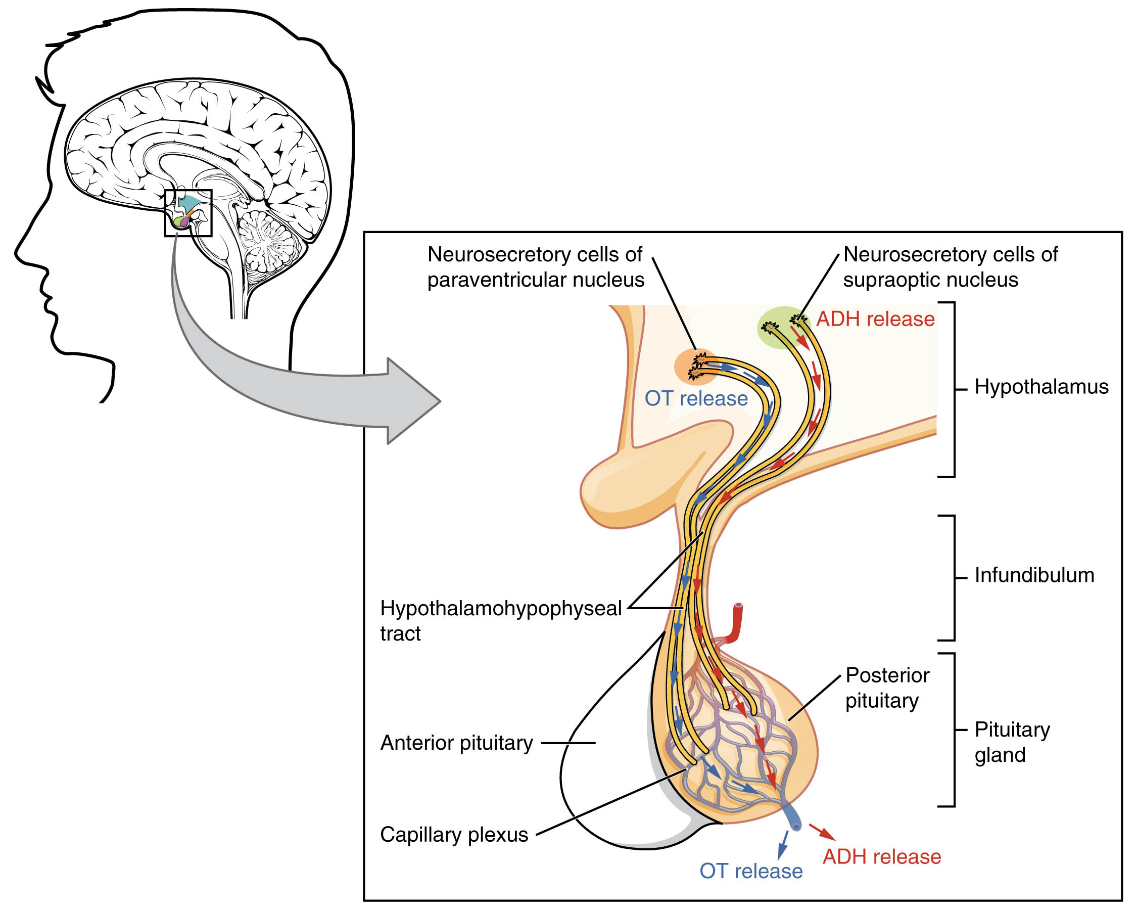
- Neurosecretory cells of paraventricular nucleus These specialized cells within the hypothalamus synthesize oxytocin, playing a key role in childbirth and lactation. They send their hormone-laden axons down the hypothalamohypophyseal tract to the posterior pituitary for storage and release.
- Neurosecretory cells of supraoptic nucleus Located in the hypothalamus, these cells produce antidiuretic hormone (ADH) to regulate water balance in the body. Their axons extend through the same tract, delivering ADH to the posterior pituitary for eventual release into the bloodstream.
- Hypothalamus The hypothalamus is a critical region that links the nervous system to the endocrine system via hormone production. It oversees the synthesis of OT and ADH, directing their transport to the pituitary gland.
- ADH release This process involves the secretion of antidiuretic hormone into the bloodstream from the posterior pituitary. ADH helps the kidneys reabsorb water, maintaining proper hydration levels and blood pressure.
- OT release Oxytocin is released from the posterior pituitary into circulation, facilitating uterine contractions during labor and milk ejection during breastfeeding. This hormone also influences social bonding and stress responses.
- Hypothalamohypophyseal tract This neural pathway transports hormones from the hypothalamus to the posterior pituitary. It consists of axons from neurosecretory cells, ensuring efficient hormone delivery.
- Infundibulum The infundibulum acts as a stalk connecting the hypothalamus to the pituitary gland. It provides a structural and functional link, allowing hormone transport through its neural connections.
- Posterior pituitary The posterior pituitary stores and releases OT and ADH into the bloodstream via the capillary plexus. It functions as an extension of the hypothalamus rather than a gland that produces hormones itself.
- Pituitary gland Comprising anterior and posterior lobes, the pituitary gland is often called the “master gland” due to its regulation of multiple hormones. The posterior portion specifically handles OT and ADH storage and release.
- Anterior pituitary This lobe produces and secretes its own hormones, such as thyroid-stimulating hormone (TSH) and adrenocorticotropic hormone (ACTH). It contrasts with the posterior pituitary by relying on a different regulatory mechanism involving the capillary plexus.
- Capillary plexus The capillary plexus is a network of blood vessels in the posterior pituitary that facilitates hormone release into the bloodstream. It ensures efficient distribution of OT and ADH to target organs.
Anatomical Overview of the Posterior Pituitary
The posterior pituitary is an essential component of the endocrine system, working in tandem with the hypothalamus to maintain hormonal balance. This section delves into its structure and function, providing a clear understanding of its role.
- The posterior pituitary does not synthesize hormones but serves as a storage depot for OT and ADH produced by the hypothalamus.
- Neurosecretory cells in the paraventricular and supraoptic nuclei extend their axons through the hypothalamohypophyseal tract to deliver these hormones.
- The infundibulum supports this connection, acting as a conduit for neural signals and hormone transport.
- Once released, OT and ADH enter the capillary plexus, which disperses them into the systemic circulation to regulate various physiological processes.
Physiological Role of Hormones in the Posterior Pituitary
Hormones released by the posterior pituitary have significant effects on the body, influencing key physiological functions. This part explores their mechanisms and importance.
- Oxytocin (OT) promotes uterine contractions during childbirth and stimulates milk let-down in lactating individuals.
- Antidiuretic hormone (ADH), also known as vasopressin, enhances water reabsorption in the kidneys, preventing dehydration.
- These hormones are released in response to specific neural signals from the hypothalamus, ensuring timely physiological adjustments.
- The balance of OT and ADH is critical for maintaining homeostasis, particularly in fluid regulation and reproductive health.
Structural Connection Between Hypothalamus and Pituitary Gland
The relationship between the hypothalamus and pituitary gland is a fascinating example of neuroendocrinology. This section examines their anatomical and functional integration.
- The hypothalamohypophyseal tract forms a direct link, allowing efficient hormone transport from the hypothalamus to the posterior pituitary.
- The infundibulum provides structural support, anchoring the pituitary gland beneath the brain.
- This connection enables rapid hormonal responses to changes in the internal environment, such as dehydration or labor onset.
- The capillary plexus further enhances this system by facilitating hormone distribution into the bloodstream.
Clinical Relevance and Hormonal Regulation
Understanding the posterior pituitary’s role can shed light on various health conditions related to hormonal imbalances. This section provides insight into its clinical significance.
- Dysfunction in ADH release can lead to conditions like diabetes insipidus, characterized by excessive urination and thirst.
- Insufficient OT production may affect lactation or uterine function, impacting postpartum recovery.
- The anterior pituitary’s distinct role in releasing hormones like TSH, which stimulates the thyroid to produce T3 and T4, complements the posterior pituitary’s functions.
- Regular monitoring of pituitary function is essential for diagnosing and managing endocrine disorders.
The posterior pituitary’s integration with the hypothalamus exemplifies the body’s intricate regulatory systems. By storing and releasing OT and ADH, it ensures critical processes like water balance and reproduction are effectively managed. This anatomical and physiological overview provides a solid foundation for further study and appreciation of endocrine health.

