The female breast is a complex and fascinating organ, crucial for lactation and a significant symbol of femininity. This article delves into the intricate anatomy of the breast, elucidating its various components as depicted in the diagram, and providing a comprehensive overview of its structure and function. Understanding breast anatomy is fundamental for recognizing potential health concerns and appreciating the physiological processes involved in milk production.
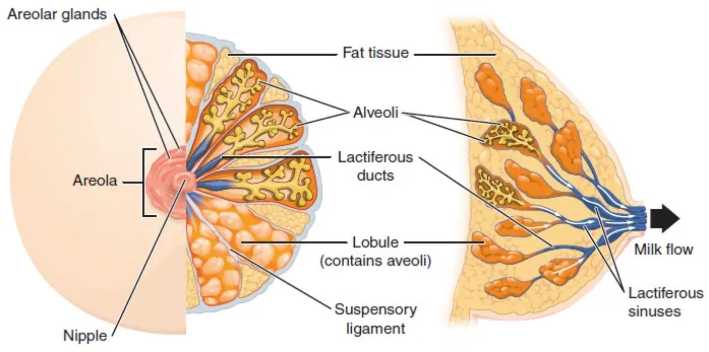
Nipple: The nipple is a raised projection located at the center of the areola. It contains several small openings through which milk is discharged during lactation, serving as the primary exit point for breast milk.
Areola: The areola is the pigmented circular area surrounding the nipple. This region often darkens during pregnancy and contains small sebaceous glands called Montgomery glands, which secrete an oily substance to lubricate and protect the nipple and areola during breastfeeding.
Areolar glands: These are also known as Montgomery glands, located within the areola. They produce a protective, lubricating substance that helps keep the nipple and areola soft and supple, which is particularly important during breastfeeding to prevent chapping and infection.
Fat tissue: Adipose tissue, or fat tissue, constitutes a significant portion of the breast’s volume and gives the breast its characteristic size and shape. It surrounds and supports the glandular tissue, providing cushioning and insulation.
Alveoli: These are tiny, sac-like structures within the breast lobules where milk is produced. Each alveolus is lined with lactocytes, specialized cells that synthesize and secrete milk in response to hormonal signals during lactation.
Lactiferous ducts: These are a network of fine tubes that transport milk from the alveoli to the lactiferous sinuses, and eventually to the nipple. As milk is produced in the alveoli, it flows through these ducts, converging into larger ducts closer to the nipple.
Lobule (contains alveoli): A lobule is a cluster of alveoli, forming the basic functional unit of the mammary gland responsible for milk production. The breast contains numerous lobules, all interconnected by a system of ducts, which collectively contribute to the milk supply.
Lactiferous sinuses: These are widened areas of the lactiferous ducts located just behind the nipple. They serve as temporary reservoirs for milk before it is expelled from the nipple during breastfeeding.
Suspensory ligament: Also known as Cooper’s ligaments, these strong, fibrous connective tissues extend from the deep fascia over the pectoralis major muscle through the breast tissue to the skin. They provide support to the breast, helping to maintain its shape and position.
The female breast is a marvel of biological engineering, primarily designed for the vital process of lactation. Its intricate internal structure allows for the efficient production and delivery of milk, providing essential nourishment to infants. Beyond its physiological role, the breast is also a significant anatomical feature influenced by hormonal fluctuations throughout a woman’s life, undergoing changes during puberty, menstrual cycles, pregnancy, and menopause.
Understanding the internal architecture of the breast is crucial for several reasons. For healthcare professionals, it underpins the diagnosis and treatment of various breast conditions, from benign lumps to more serious concerns like breast cancer. For individuals, knowledge of normal breast anatomy can empower them to perform self-examinations and recognize any unusual changes that warrant medical attention. The breast is composed of:
- Glandular tissue (lobules and ducts)
- Fatty tissue
- Fibrous connective tissue (ligaments)
- Nerves, blood vessels, and lymphatic vessels
During pregnancy, hormonal shifts, particularly increases in estrogen and progesterone, stimulate the development of the glandular tissue. The alveoli mature, and the ducts proliferate in preparation for milk production. Following childbirth, the hormone prolactin triggers milk synthesis within the alveoli, while oxytocin facilitates the milk ejection reflex, causing milk to flow through the lactiferous ducts and sinuses to the nipple.
The delicate balance of these structures and their coordinated function ensure the breast can fulfill its primary biological role. Variations in breast size and shape are common among individuals and are largely determined by the amount of fat tissue present, as the glandular tissue itself is relatively consistent in volume across most women. Regular breast health practices, including clinical examinations and imaging, rely heavily on a thorough understanding of this detailed anatomy.
In conclusion, the female breast is a dynamic and essential organ with a complex anatomical design perfectly suited for its reproductive function. From the milk-producing alveoli to the supporting suspensory ligaments and the network of ducts that transport milk, each component plays a critical role. Appreciating this intricate structure not only enhances our understanding of human biology but also underscores the importance of breast health awareness and regular medical check-ups to maintain overall well-being.

