The cranial nerves are a fundamental component of the peripheral nervous system, consisting of 12 pairs that emerge directly from the brain and brainstem. These nerves play crucial roles in sensory perception, motor control, and autonomic functions, facilitating everything from vision and hearing to taste and visceral regulation. This article explores their anatomical arrangement as seen in an inferior view of the brain, highlighting their origins, pathways, and physiological significance to provide a comprehensive understanding of how they integrate with the central nervous system.
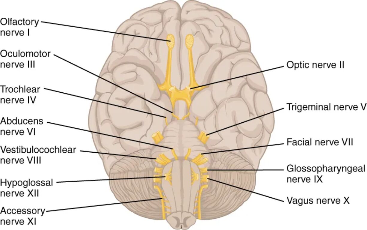
Labeled Parts of the Cranial Nerves
Olfactory nerve I The olfactory nerve (CN I) is responsible for the sense of smell, transmitting sensory information from the nasal mucosa to the olfactory bulb in the brain. It consists of numerous small filaments that pass through the cribriform plate of the ethmoid bone, making it unique as the only cranial nerve that directly connects to the cerebral cortex without synapsing in the thalamus.
Oculomotor nerve III The oculomotor nerve (CN III) controls most of the eye’s extrinsic muscles, enabling movements such as elevation, depression, adduction, and medial rotation of the eyeball. It also innervates the levator palpebrae superioris for eyelid elevation and carries parasympathetic fibers to the ciliary muscle and sphincter pupillae for accommodation and pupillary constriction.
Optic nerve II The optic nerve (CN II) conveys visual information from the retina to the brain, forming the pathway for sight through the optic chiasm and tracts. As a direct extension of the central nervous system, it is surrounded by meninges and can be affected by increased intracranial pressure, leading to conditions like papilledema.
Trochlear nerve IV The trochlear nerve (CN IV) innervates the superior oblique muscle of the eye, which primarily depresses and intorts the eyeball. It has the longest intracranial course among the cranial nerves and decussates before exiting the brainstem, making it vulnerable to trauma or compression.
Trigeminal nerve V The trigeminal nerve (CN V) is the largest cranial nerve, providing sensory innervation to the face, mouth, and nasal cavity, as well as motor control to the muscles of mastication. It divides into three branches—ophthalmic, maxillary, and mandibular—each serving distinct regions, and its sensory ganglia can be involved in conditions like trigeminal neuralgia.
Abducens nerve VI The abducens nerve (CN VI) supplies the lateral rectus muscle, facilitating abduction of the eyeball. Its long course along the base of the skull makes it susceptible to damage from increased intracranial pressure or fractures, often resulting in diplopia or strabismus.
Vestibulocochlear nerve VIII The vestibulocochlear nerve (CN VIII) handles hearing and balance, with its cochlear branch transmitting auditory signals and the vestibular branch conveying equilibrium information from the inner ear. It enters the brainstem at the pontomedullary junction, and dysfunction can lead to vertigo, tinnitus, or hearing loss.
Facial nerve VII The facial nerve (CN VII) controls the muscles of facial expression, provides taste sensation to the anterior two-thirds of the tongue, and supplies parasympathetic innervation to salivary and lacrimal glands. It travels through the facial canal in the temporal bone, where it can be compressed in conditions like Bell’s palsy.
Glossopharyngeal nerve IX The glossopharyngeal nerve (CN IX) innervates the stylopharyngeus muscle for swallowing and provides sensory input from the posterior third of the tongue, pharynx, and carotid body. It also carries parasympathetic fibers to the parotid gland, playing a role in salivation and baroreceptor reflexes.
Vagus nerve X The vagus nerve (CN X) is the longest cranial nerve, extending from the brainstem to the abdomen, regulating parasympathetic functions in the heart, lungs, and digestive tract. It mediates the gag reflex, voice production via laryngeal innervation, and visceral sensory feedback, influencing autonomic homeostasis.
Hypoglossal nerve XII The hypoglossal nerve (CN XII) provides motor innervation to the intrinsic and extrinsic muscles of the tongue, essential for speech, swallowing, and mastication. It exits the medulla through the hypoglossal canal, and unilateral damage can cause tongue deviation toward the affected side.
Accessory nerve XI The accessory nerve (CN XI) has cranial and spinal roots; the cranial root joins the vagus for pharyngeal and laryngeal innervation, while the spinal root supplies the sternocleidomastoid and trapezius muscles for head and shoulder movements. It is unique in having contributions from both the brainstem and upper cervical spinal cord.
Anatomy of the Cranial Nerves: Origins and Pathways
The cranial nerves originate from specific nuclei within the brainstem, with their roots visible in the inferior view of the brain as depicted in the image. This perspective allows for a clear observation of how they emerge in a sequential manner from the forebrain, midbrain, pons, and medulla oblongata.
- Forebrain origins: The olfactory nerve (I) and optic nerve (II) arise from the telencephalon and diencephalon, respectively, differing from the brainstem-emerging nerves.
- Midbrain connections: Nerves like the oculomotor (III) and trochlear (IV) exit from the midbrain, crossing paths to innervate contralateral eye muscles.
- Pontine attachments: The trigeminal (V), abducens (VI), facial (VII), and vestibulocochlear (VIII) nerves attach at the pons, forming a cluster that handles facial sensation, eye movement, expression, and auditory-vestibular functions.
- Medullary roots: The glossopharyngeal (IX), vagus (X), accessory (XI), and hypoglossal (XII) nerves emerge from the medulla, extending to control swallowing, autonomic regulation, and tongue movements.
- Pathways through foramina: Each nerve passes through specific skull base openings, such as the optic canal for CN II or the jugular foramen for CN IX, X, and XI, which can be sites of compression in pathologies.
Physiological Functions of the Cranial Nerves
Each cranial nerve serves specialized functions, categorized as sensory, motor, or mixed, contributing to overall neurological integrity. Understanding these roles is essential for assessing neurological exams and diagnosing disorders.
- Sensory nerves: Purely sensory nerves like CN I (olfaction), CN II (vision), and CN VIII (hearing and balance) relay environmental and internal stimuli to the brain without motor components.
- Motor nerves: Nerves such as CN III, IV, VI (eye movements), CN XI (neck and shoulder), and CN XII (tongue) are primarily motor, ensuring precise voluntary movements.
- Mixed nerves: CN V (facial sensation and mastication), CN VII (facial expression, taste, and secretion), CN IX (pharyngeal sensation and salivation), and CN X (visceral control and sensation) combine sensory and motor fibers, often with parasympathetic elements.
- Parasympathetic contributions: CN III, VII, IX, and X carry parasympathetic fibers from the brainstem nuclei, regulating pupil size, glandular secretion, and organ functions like heart rate modulation via the vagus.
- Integration with CNS: These nerves synapse with brainstem nuclei, influencing reflexes such as the pupillary light reflex (CN II and III) or the corneal reflex (CN V and VII).
Clinical Relevance and Assessment of Cranial Nerves
Evaluating cranial nerve function is a cornerstone of neurological examinations, often revealing underlying pathologies. Common tests include assessing smell for CN I, visual fields for CN II, and eye movements for CN III, IV, and VI.
- Common disorders: Damage to CN VII can cause facial paralysis, while CN V involvement may lead to intense facial pain; CN X dysfunction affects swallowing and voice.
- Diagnostic tools: Imaging like MRI visualizes nerve roots in the inferior brain view, aiding in identifying tumors or vascular compressions.
- Reflex testing: The jaw jerk reflex tests CN V, and the gag reflex evaluates CN IX and X, providing insights into brainstem integrity.
- Systemic impacts: Autonomic dysregulation from CN X can influence cardiovascular and gastrointestinal systems, linking neurology to internal medicine.
Evolutionary and Developmental Aspects
Cranial nerves have evolved from primitive segmental nerves in vertebrates, adapting to complex sensory and motor needs in humans. During embryogenesis, they develop from neural crest cells and placodes, with their arrangement conserved across species.
- Phylogenetic origins: Nerves like CN X reflect ancient gill arch innervations, now adapted for visceral control in air-breathing mammals.
- Developmental anomalies: Congenital defects, such as absent CN I in Kallmann syndrome, highlight the importance of proper neural migration.
- Comparative anatomy: In humans, the inferior view shows a compact brainstem exit, contrasting with elongated structures in some animals.
In summary, the 12 pairs of cranial nerves represent a sophisticated network bridging the brain to the body, essential for sensory-motor coordination and autonomic balance. Their anatomical layout, as illustrated in the inferior brain view, underscores the precision of human neuroanatomy, offering insights into both health and disease management.

