Retinal disparity plays a crucial role in how the human visual system interprets depth and three-dimensional space from two-dimensional retinal images. This fundamental concept in visual perception highlights the differences in the positions of images on the retinas of each eye, enabling the brain to compute distance and create a sense of depth.
Labels in the Diagram
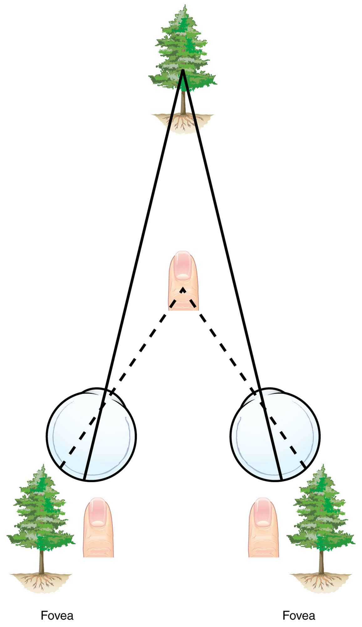
Fovea The fovea is the central region of the retina responsible for high-acuity vision, where cone cells are densely packed to provide sharp, detailed color perception. In this diagram, the fovea labels indicate the points on the retinas where the distant object’s image falls, demonstrating how fixation on a far point affects the projection of closer objects.
Fovea (Left Retina) This fovea represents the high-resolution area in the left eye’s retina, shown with the image of the distant tree centered on it. The offset position of the finger’s image relative to this fovea illustrates the horizontal disparity used by the brain for stereopsis.
Fovea (Right Retina) Similarly, the fovea in the right eye’s retina captures the central image of the distant tree, while the finger appears displaced to the side. This disparity between the two foveal projections is essential for binocular vision, allowing precise depth cues without eye movement.
The Basics of Retinal Disparity
Retinal disparity arises from the slight separation between the two eyes, known as interocular distance, which causes objects at varying depths to project onto different retinal locations. This phenomenon is the foundation of stereoscopic vision, where the brain fuses the two slightly different images into a single three-dimensional percept.
Understanding this process begins with the anatomy of the eye. Light rays from an object pass through the cornea and lens, inverting the image on the retina. In the diagram, the top tree represents a distant fixation point, its image landing on the fovea of both retinas, while the closer finger creates disparate images.
- Interocular Distance: Typically around 6-7 cm in adults, this separation ensures each eye views the world from a unique angle.
- Horizontal Disparity: The key metric for depth, measured as the difference in horizontal positions on the retinas.
- Convergence and Accommodation: These work in tandem with disparity; convergence angles the eyes inward for near objects, while accommodation adjusts lens focus.
The brain processes these disparities in the visual cortex, specifically in areas like V1 and V2, where neurons tuned to specific disparity levels fire to signal depth.
Anatomical Structures Involved in Depth Perception
The retina, a thin layer of neural tissue at the back of the eye, contains photoreceptors that convert light into electrical signals for the brain. The fovea, as highlighted in the diagram, is a small pit in the macula lutea with the highest concentration of cones, enabling detailed vision essential for perceiving fine disparities.
Beyond the retina, the optic nerves carry signals to the lateral geniculate nucleus and then to the visual cortex. Here, binocular cells integrate inputs from both eyes to detect disparities.
- Photoreceptors: Rods for low-light vision and cones for color and detail; cones dominate the fovea.
- Optic Chiasm: Where nasal fibers cross, ensuring contralateral processing for binocular integration.
- Visual Cortex Layers: Layer 4 receives inputs, while higher layers compute complex features like depth.
Disorders affecting these structures, such as amblyopia, can impair disparity detection, leading to reduced depth perception.
Physiological Mechanisms of Binocular Vision
Binocular vision relies on the precise alignment and coordination of both eyes to achieve fusion and stereopsis. When objects are at different distances, the resulting retinal disparity provides quantitative depth information, with crossed disparity for near objects and uncrossed for far ones.
This mechanism evolved to aid in tasks like predation and navigation. The diagram simplifies this by showing a far tree on the foveas and a near finger offset, mimicking real-world viewing.
- Stereopsis: The perception of depth from disparity alone, testable with random-dot stereograms.
- Panum’s Fusional Area: The range of disparities where images fuse without diplopia.
- Vergence Movements: Eye rotations that align images on corresponding retinal points.
Neural pathways involve magnocellular and parvocellular streams, with the former handling motion and depth.
Applications and Implications in Everyday Life
In daily activities, retinal disparity helps us judge distances for actions like catching a ball or driving. Without it, as in monocular vision, we rely on secondary cues like motion parallax or shading.
The diagram underscores why strabismus, or eye misalignment, disrupts this process, potentially causing double vision.
- Virtual Reality: Systems exploit disparity to create immersive 3D environments.
- Ophthalmic Testing: Tools like stereoscopes assess disparity sensitivity for diagnosing visual impairments.
- Evolutionary Perspective: Primates with forward-facing eyes benefit from enhanced depth for arboreal life.
Advancements in neuroimaging reveal how disparity-selective neurons cluster in the brain, aiding research into visual prosthetics.
Challenges and Variations in Depth Perception
Individual differences in interocular distance affect disparity magnitude, with larger separations enhancing depth sensitivity at greater distances. Age-related changes, such as reduced lens flexibility, can indirectly impact disparity processing by altering focus cues.
The diagram’s simplified model assumes perfect alignment, but in reality, factors like head tilt or asymmetrical eye positions introduce complexities.
- Aniseikonia: Unequal image sizes between eyes, complicating fusion.
- Suppression: In strabismus, the brain ignores one eye’s input to avoid confusion.
- Training Effects: Visual therapy can improve disparity detection in certain conditions.
Understanding these variations is vital for fields like optometry and neurology.
In summary, retinal disparity transforms flat retinal images into a rich, depth-filled world, as elegantly illustrated in this diagram. By appreciating the roles of the fovea and binocular integration, we gain insight into the marvels of human vision and its underlying physiology.

