Neurulation is a pivotal embryonic process that marks the initial formation of the central nervous system (CNS) and lays the groundwork for the axial skeleton. This intricate series of events transforms a flat sheet of embryonic tissue into the neural tube, which will eventually develop into the brain and spinal cord. The diagram provided illustrates the sequential stages of neurulation, from the initial thickening of the neural plate to the closure of the neural tube and the differentiation of surrounding structures. Understanding neurulation is fundamental to comprehending the origins of the nervous system and the potential implications of developmental anomalies.
Key Structures and Stages in Neurulation
Neural plate border: This is the region where the neuroectodermal tissue of the neural plate meets the surrounding ectoderm. Cells at this border play a crucial role in forming the neural crest.
Neural plate: A thickened, flat plate of ectoderm that forms in response to signals from the underlying notochord. It is the precursor to the entire central nervous system.
Ectoderm: The outermost of the three primary germ layers of the embryo. It gives rise to the epidermis, nervous system, and other structures.
Mesoderm: One of the three primary germ layers, located between the ectoderm and endoderm. It forms various tissues including muscle, bone, connective tissue, and the notochord.
Notochord: A flexible, rod-like structure derived from the mesoderm, located ventral to the neural plate/tube. It induces the formation of the neural plate and later contributes to the nucleus pulposus of intervertebral discs.
Convergence: This term refers to the process where cells migrate towards the midline, causing the neural plate to narrow and lengthen. This movement is essential for the subsequent folding of the neural plate.
Neural crest: A transient group of cells that delaminates from the neural plate border during neural tube closure. These highly migratory cells differentiate into a wide array of cell types, forming components of the peripheral nervous system and many other tissues.
Neural crest cells (form components of the PNS): These are the cells that originate from the neural crest and differentiate into a diverse range of cell types. They form a significant portion of the peripheral nervous system, including sensory neurons, autonomic ganglia, and Schwann cells.
Neural tube: The hollow tube formed by the invagination and closure of the neural plate. It is the embryonic precursor to the brain and spinal cord.
Epidermis: The outermost layer of the skin, derived from the ectoderm. Following neural tube closure, the remaining surface ectoderm will form the epidermis.
Spinal ganglion: Also known as dorsal root ganglia, these are clusters of sensory neuron cell bodies located along the spinal cord. They are derived from neural crest cells.
Somites (mesoderm-derived): Blocks of mesoderm that form along the neural tube. They are precursors to the axial skeleton (vertebrae and ribs), skeletal muscle, and dermis of the skin.
The Sequential Process of Neurulation
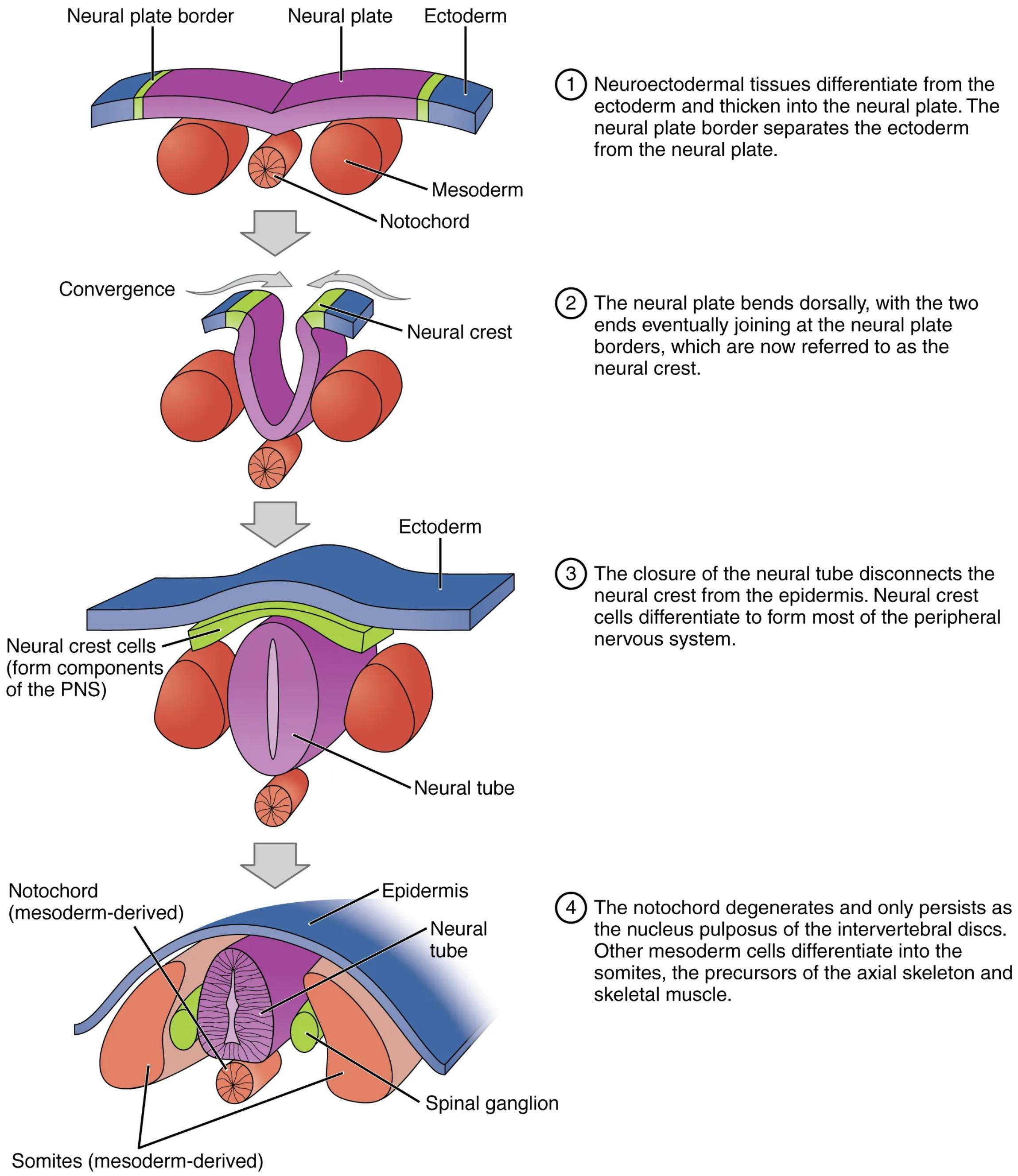
The process begins with the differentiation of neuroectodermal tissues from the ectoderm, leading to the formation of the neural plate. This flat structure is separated from the surrounding ectoderm by the neural plate border. The underlying notochord provides crucial inductive signals that initiate and guide this transformation.
Subsequently, the neural plate undergoes a process of bending and folding dorsally. The edges of the neural plate elevate to form neural folds, with a neural groove developing centrally. These neural folds eventually meet at the midline and fuse, a process referred to as neural tube closure. This fusion typically begins in the cervical region and proceeds bidirectionally, both rostrally and caudally.
Upon the closure of the neural tube, the neural crest cells, which originated from the neural plate border, disconnect from the overlying epidermis. These remarkable cells then embark on extensive migrations throughout the embryo, differentiating into a vast array of cell types. These include neurons and glia of the peripheral nervous system, chromaffin cells of the adrenal medulla, melanocytes, and components of the craniofacial skeleton. Meanwhile, the notochord, having completed its inductive role, largely degenerates, with remnants persisting as the nucleus pulposus of the intervertebral discs. Concurrently, the mesoderm adjacent to the neural tube segments into somites, which will give rise to the axial skeleton and skeletal muscles.
- Disruptions in neural tube closure can lead to severe birth defects known as neural tube defects (NTDs), such as spina bifida and anencephaly.
This orchestrated sequence of cellular movements and differentiations is fundamental for the proper development of the central and peripheral nervous systems, as well as essential musculoskeletal structures.
Conclusion
Neurulation is an extraordinary example of coordinated embryonic development, transforming simple germ layers into the complex beginnings of the nervous system and skeleton. The diagram lucidly illustrates the critical steps involved, from the induction of the neural plate to the formation and closure of the neural tube, and the subsequent differentiation of neural crest cells and somites. A thorough understanding of neurulation is essential not only for appreciating normal embryonic development but also for recognizing the origins of various congenital anomalies.

