Understanding Blood Composition: Hematocrit Levels in Normal, Anemic, and Polycythemic States
Blood is a vital fluid in the human body, responsible for transporting oxygen, nutrients, and waste products while also playing a key role in immune defense and clotting. This diagram illustrates the centrifuged components of blood, highlighting the differences between normal blood and conditions like anemia and polycythemia through visual representations of plasma, buffy coat, and hematocrit. By examining these layers, healthcare professionals can assess red blood cell volume and diagnose various disorders.
Key Components of Blood as Shown in the Diagram
The diagram depicts blood separated into distinct layers after centrifugation, providing a clear view of its composition.
Plasma:
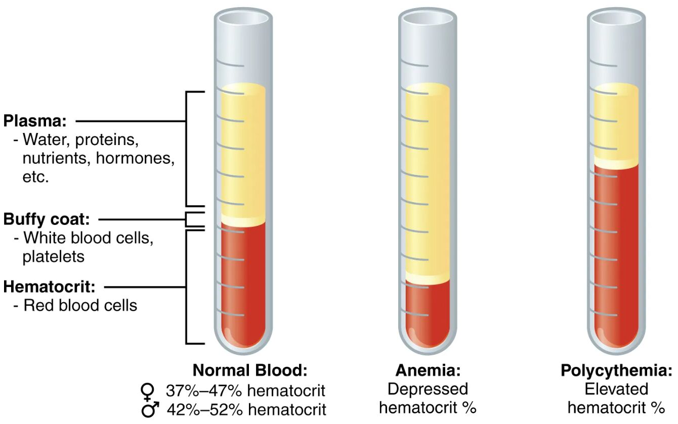
Plasma is the liquid portion of blood, comprising about 55% of total blood volume, and serves as the medium for suspending blood cells and transporting essential substances. It contains water as its primary component, along with proteins such as albumin and globulins, nutrients like glucose and amino acids, hormones including insulin and cortisol, and electrolytes that maintain osmotic balance and pH levels.
Buffy coat:
The buffy coat is a thin, whitish layer that forms between the plasma and the red blood cells during centrifugation, representing less than 1% of blood volume. It consists primarily of white blood cells, which are crucial for immune responses against infections, and platelets, which are essential for blood clotting and wound healing.
Hematocrit:
Hematocrit refers to the percentage of blood volume occupied by red blood cells, typically measured after centrifugation to separate cellular components. It is a critical indicator of oxygen-carrying capacity, with abnormal levels signaling conditions like dehydration or blood disorders.
Normal Blood:
Normal blood exhibits a balanced composition where the hematocrit falls within standard ranges, ensuring efficient oxygen transport and overall homeostasis. For females, this is typically 37%-47%, while for males, it ranges from 42%-52%, reflecting physiological differences in body composition and hormone influences.
Anemia:
Anemia is characterized by a depressed hematocrit percentage, indicating a reduced number or volume of red blood cells. This condition can lead to symptoms such as fatigue and shortness of breath due to diminished oxygen delivery to tissues.
Polycythemia:
Polycythemia features an elevated hematocrit percentage, signifying an increased concentration of red blood cells. This can result from chronic hypoxia or other underlying causes, potentially leading to increased blood viscosity and cardiovascular strain.
The Physiological Role of Blood Composition
Blood composition is fundamental to maintaining bodily functions, as it ensures the delivery of oxygen and nutrients while removing waste. Variations in these components, as depicted in the diagram, can provide insights into health status and guide diagnostic processes.
The plasma layer, being the largest component, acts as a solvent for various solutes and facilitates the transport of clotting factors and antibodies. Its yellowish hue in the diagram comes from dissolved proteins and lipids. The buffy coat, though small, is packed with leukocytes like neutrophils and lymphocytes, which combat pathogens, and thrombocytes that prevent excessive bleeding. Hematocrit, shown as the red layer, directly correlates with hemoglobin levels, influencing blood’s ability to bind and release oxygen via the oxyhemoglobin dissociation curve.
In normal blood, the proportions allow for optimal flow through capillaries without excessive thickness or dilution. This balance is regulated by hormones such as erythropoietin from the kidneys, which stimulates red blood cell production in the bone marrow. Disruptions in this equilibrium, as seen in the anemic and polycythemic samples, highlight how environmental factors, nutrition, and genetics interplay in hematopoiesis.
- Plasma Functions: Transports hormones like triiodothyronine (T3) and thyroxine (T4) from the thyroid gland, which regulate metabolism; carries fibrinogen for clot formation.
- Buffy Coat Contributions: White blood cells include basophils that release histamine in allergic responses; platelets aggregate at injury sites to form plugs.
- Hematocrit Implications: Normal ranges prevent issues like tissue hypoxia or thrombosis; measurements are part of complete blood counts (CBC) in clinical settings.
Anemia: Causes, Symptoms, and Management
Anemia affects millions worldwide, often stemming from nutritional deficiencies or chronic diseases, and is visually represented by a smaller red cell layer in the diagram. Early detection through hematocrit assessment can prevent complications like heart failure.
Common causes include iron deficiency, where inadequate dietary iron impairs hemoglobin synthesis, leading to microcytic anemia. Vitamin B12 or folate deficiencies result in megaloblastic anemia, characterized by large, immature red blood cells. Chronic conditions such as kidney disease reduce erythropoietin production, while hemolytic anemias involve premature red cell destruction due to autoimmune factors or genetic defects like sickle cell disease.
Symptoms manifest as pallor, weakness, and tachycardia, as the body compensates for low oxygen-carrying capacity. Diagnosis involves laboratory tests beyond hematocrit, including mean corpuscular volume (MCV) and reticulocyte count to classify the type.
Management strategies depend on etiology:
- Nutritional supplementation for deficiencies, such as oral iron for iron-deficiency anemia.
- Blood transfusions in severe cases to rapidly restore hematocrit.
- Treating underlying conditions, like erythropoietin analogs for renal anemia.
- Lifestyle modifications, including a diet rich in leafy greens and lean meats to boost iron absorption.
Prognosis is generally favorable with timely intervention, though chronic forms require ongoing monitoring to avoid organ damage.
Polycythemia: Understanding Elevated Red Blood Cell Mass
Polycythemia, illustrated by an enlarged hematocrit layer, increases blood viscosity and poses risks like stroke or heart attack. It can be primary, due to bone marrow overproduction, or secondary, triggered by external factors.
Primary polycythemia vera arises from JAK2 gene mutations, leading to uncontrolled erythroid proliferation independent of erythropoietin. Secondary forms often result from chronic hypoxia in high-altitude dwellers or smokers, stimulating excessive red cell production. Other causes include tumors secreting erythropoietin or dehydration concentrating blood components.
Clinical features include ruddy complexion, headaches, and pruritus after bathing, stemming from hyperviscosity. Diagnostic criteria involve elevated hemoglobin, hematocrit above 49% in men or 48% in women, and bone marrow biopsy for confirmation.
Treatment aims to reduce red cell mass and prevent thrombosis:
- Phlebotomy to remove excess blood and lower hematocrit.
- Medications like hydroxyurea to suppress marrow activity in polycythemia vera.
- Aspirin for antiplatelet effects to mitigate clotting risks.
- Addressing root causes, such as smoking cessation or oxygen therapy for hypoxic patients.
Regular follow-up is essential, as untreated cases can progress to myelofibrosis or acute leukemia.
Clinical Significance and Diagnostic Applications
Centrifuged blood analysis, as shown, is a cornerstone in hematology for evaluating disorders. It provides a quick visual assessment before advanced testing.
In practice, hematocrit is measured via automated analyzers, correlating with manual spins in the diagram. Deviations prompt further investigation, like serum ferritin for anemia or erythropoietin levels for polycythemia. This approach aids in differentiating relative from absolute polycythemia, where plasma volume changes mimic true increases.
Preventive measures include balanced nutrition, hydration, and avoiding tobacco to maintain normal composition. Research continues into genetic therapies for inherited forms, promising targeted treatments.
Blood composition diagrams like this one underscore the delicate balance required for health, reminding us that even small shifts in layers can signal significant pathologies. By understanding these visuals, one gains appreciation for the body’s intricate regulatory systems, from hormonal controls to cellular responses, ensuring vitality across diverse physiological demands.

