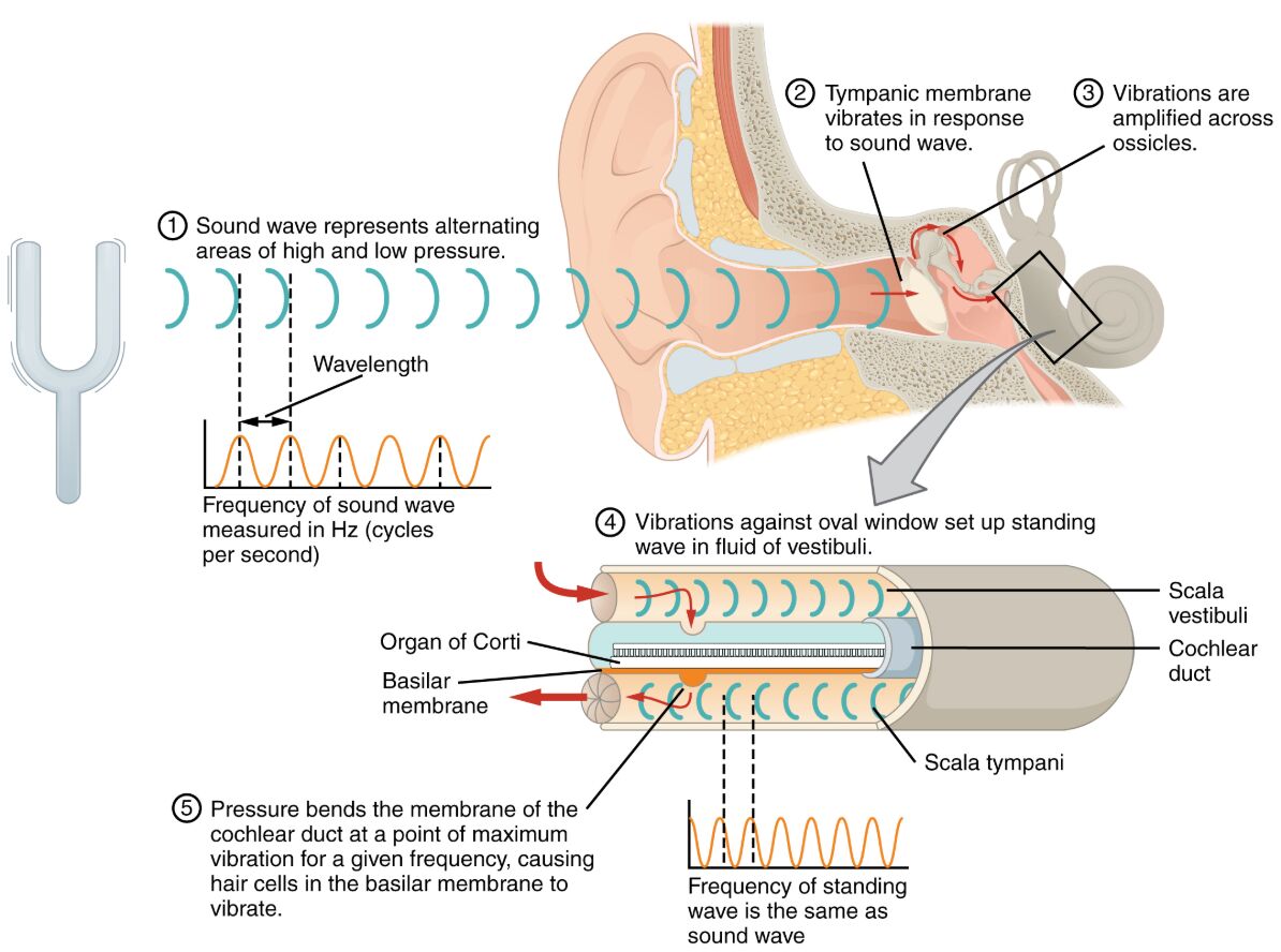
The journey of sound from the environment to the inner ear is a fascinating process that begins with the vibration of the tympanic membrane and culminates in the cochlea’s fluid dynamics. This image illustrates how sound waves are amplified through the ossicles—malleus, incus, and stapes—and transformed into pressure waves within the scala vestibuli and scala tympani, enabling auditory perception. This article delves into the anatomical and physiological mechanisms behind this transmission, offering a comprehensive understanding of how the ear converts sound into meaningful signals.
Labeled Parts of Sound Wave Transmission
Tympanic membrane The tympanic membrane, or eardrum, is a thin, flexible structure that vibrates when struck by sound waves entering the ear canal. These vibrations are the first step in the auditory process, transferring energy to the ossicles for further amplification.
Malleus The malleus, the outermost of the three ossicles, is attached to the tympanic membrane and transmits its vibrations to the incus. Its hammer-like shape ensures efficient energy transfer, initiating the mechanical amplification of sound.
Incus The incus, or anvil, is the middle ossicle that receives vibrations from the malleus and passes them to the stapes. Its pivotal role in the ossicular chain helps maintain the continuity and precision of sound wave movement.
Stapes The stapes, the smallest bone in the human body, connects to the oval window and transfers amplified vibrations from the incus to the inner ear. Its footplate movement against the oval window generates pressure waves critical for cochlear function.
Oval window The oval window is a membrane-covered opening between the middle ear and the cochlea that receives vibrations from the stapes. It transmits these vibrations into the fluid-filled scala vestibuli, initiating the process of sound transduction.
Scala vestibuli The scala vestibuli is an upper fluid-filled chamber of the cochlea that carries pressure waves from the oval window toward the apex. These waves stimulate the cochlear partition, contributing to the perception of sound pitch and intensity.
Scala tympani The scala tympani is a lower fluid-filled chamber of the cochlea that extends from the apex to the round window, completing the pressure wave pathway. It dissipates excess pressure, ensuring the cochlea’s fluid dynamics remain balanced during sound processing.
Round window The round window is a flexible membrane at the base of the scala tympani that relieves pressure generated by sound waves in the cochlear fluid. Its movement allows the cochlea to respond to varying sound frequencies without damage.
Anatomical Overview of Sound Transmission
The transmission of sound waves to the cochlea involves a coordinated sequence of structures from the middle to the inner ear. This anatomical pathway ensures that sound is efficiently captured, amplified, and converted into neural signals.
- External to middle ear: The tympanic membrane captures sound waves, setting the ossicles—malleus, incus, and stapes—into motion.
- Ossicular amplification: The ossicles increase the force of vibrations, with the stapes delivering enhanced energy to the oval window.
- Cochlear entry: The oval window transmits these vibrations into the scala vestibuli, initiating fluid movement within the cochlea.
- Fluid dynamics: The scala tympani and scala vestibuli work together, with the round window releasing pressure to maintain cochlear function.
- Protective design: The ear’s structure minimizes energy loss, ensuring accurate sound transmission to the auditory nerve.
Physiological Functions of Sound Wave Transmission
The ear’s mechanisms transform acoustic energy into electrical signals, enabling the perception of sound across a wide range of frequencies and amplitudes. This process relies on the precise interaction of its components.
- Vibration initiation: The tympanic membrane vibrates in response to sound waves, converting air pressure changes into mechanical motion.
- Amplification process: The malleus, incus, and stapes amplify these vibrations, increasing pressure at the oval window by a factor of about 20.
- Fluid wave generation: The oval window creates pressure waves in the scala vestibuli, which travel through the cochlear fluid to stimulate hair cells.
- Pressure regulation: The scala tympani and round window manage fluid displacement, allowing the cochlea to handle complex sound patterns.
- Signal conversion: Hair cells in the cochlea translate these pressure waves into electrical impulses, sent to the brain via the auditory nerve.
Developmental and Structural Dynamics
The structures involved in sound transmission develop during embryogenesis, maturing to support hearing by early childhood. This development reflects both genetic and environmental influences on auditory function.
- Embryonic formation: The tympanic membrane and ossicles form from the first and second pharyngeal arches, establishing the middle ear.
- Ossicle maturation: The malleus, incus, and stapes develop into a functional chain, with the stapes connecting to the oval window prenatally.
- Cochlear growth: The scala vestibuli and scala tympani form as the cochlea spirals, with hair cells differentiating for sound detection.
- Pressure relief: The round window develops to accommodate fluid dynamics, ensuring cochlear integrity during sound processing.
- Postnatal refinement: The ear’s structures adapt to external sounds, with the ossicles fine-tuning their movement over time.
Clinical Relevance and Auditory Health
Understanding sound wave transmission is essential for diagnosing and managing hearing-related conditions. Clinical assessments often focus on these structures to identify dysfunction and guide treatment.
- Conductive hearing loss: Damage to the tympanic membrane or ossicles, such as from otitis media, can impede sound transmission.
- Sensorineural loss: Issues with the cochlea, including hair cell damage, may result from noise exposure or aging, affecting signal conversion.
- Otosclerosis: Abnormal bone growth around the stapes can restrict oval window movement, leading to hearing impairment.
- Diagnostic tools: Audiometry and tympanometry evaluate the function of the ossicles and cochlear fluid dynamics.
- Therapeutic options: Treatments range from hearing aids to surgical procedures like stapedectomy to restore sound conduction.
In conclusion, the transmission of sound waves to the cochlea represents a sophisticated interplay of anatomical structures and physiological processes, from the vibrating tympanic membrane to the fluid-filled scalae. This image highlights the precision with which the ear amplifies and converts sound, offering insights into both normal auditory function and potential clinical interventions. Exploring these mechanisms enhances appreciation for the ear’s role in communication and balance, underscoring the importance of maintaining its health.

