The visual system intricately maps the external world onto the retina, creating an inverted and reversed image that is faithfully preserved as it travels through the visual pathway to the cortex. This diagram illustrates how this topographic organization ensures that spatial relationships in the visual field are maintained, providing a clear representation of how the brain interprets what we see.
Retina The retina is the light-sensitive layer at the back of the eye where the visual field is initially projected as an inverted, reversed image. It contains photoreceptors that convert light into electrical signals, which are then transmitted via ganglion cells.
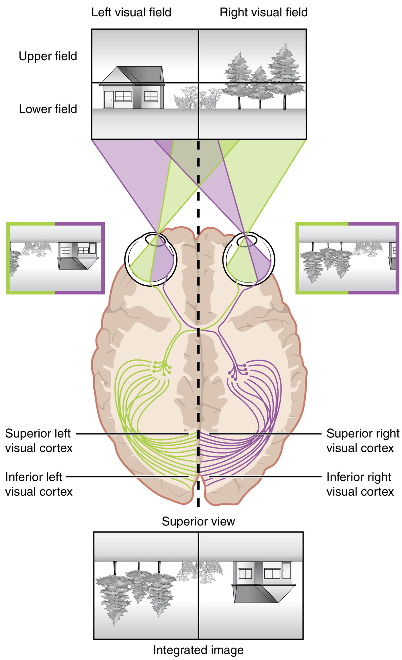
Optic nerve (II) The optic nerve (II) carries visual signals from the retina to the optic chiasm, maintaining the topographic arrangement of the retinal image. It consists of axons from retinal ganglion cells, preserving spatial information.
Optic chiasm The optic chiasm is where some optic nerve fibers cross to the opposite side, segregating nasal and temporal retinal inputs. This partial decussation ensures that each hemisphere processes the contralateral visual field.
Optic tract The optic tract transports a mix of crossed and uncrossed fibers from the optic chiasm to the lateral geniculate nucleus, retaining the topographic map. It relays the organized visual data for further processing.
Lateral geniculate nucleus (LGN) The lateral geniculate nucleus (LGN), located in the thalamus, receives input from the optic tract and organizes it into layers that preserve retinal topography. It acts as a relay station, forwarding signals to the visual cortex.
Optic radiation The optic radiation consists of fiber tracts that carry visual information from the LGN to the visual cortex, maintaining the spatial layout. These fibers fan out, ensuring the topographic map reaches the appropriate cortical areas.
Visual cortex (V1) The visual cortex (V1), located in the occipital lobe, receives the topographic map via the optic radiation and processes it into a coherent visual image. It is organized into a retinotopic map, where adjacent retinal points map to adjacent cortical areas.
Left visual field The left visual field is represented in the right hemisphere, with input from the temporal retina of the left eye and nasal retina of the right eye crossing at the optic chiasm. This arrangement allows binocular integration of the visual scene.
Right visual field The right visual field is processed by the left hemisphere, receiving input from the temporal retina of the right eye and nasal retina of the left eye after decussation. This ensures a complete representation of the visual environment.
Anatomy of the Visual Pathway
The visual pathway maintains a topographic map from the retina to the visual cortex (V1), preserving spatial relationships. This diagram outlines the anatomical structures involved in this process.
- The retina captures the inverted image through the lens system.
- The optic nerve (II) begins the journey, carrying signals to the optic chiasm.
- The optic chiasm facilitates the crossing of nasal retinal fibers.
- The optic tract and lateral geniculate nucleus (LGN) continue the relay.
- The optic radiation distributes the map to the visual cortex (V1).
- The left visual field and right visual field are segregated for hemispheric processing.
- The pathway’s organization reflects the retina’s point-to-point projection.
Physiology of Topographic Mapping
The topographic mapping ensures that the retina’s inverted image is accurately represented in the visual cortex (V1). This diagram illustrates the physiological process of visual signal transmission.
- The retina’s photoreceptors detect light and send signals through ganglion cells.
- The optic nerve (II) preserves the spatial arrangement as it reaches the optic chiasm.
- The optic chiasm reorganizes fibers, with nasal inputs crossing to the opposite side.
- The optic tract and lateral geniculate nucleus (LGN) maintain this organization.
- The optic radiation projects the map to the visual cortex (V1) in a retinotopic manner.
- The left visual field and right visual field are integrated for binocular vision.
- This mapping allows the brain to reconstruct the upright visual scene.
Role of the Optic Chiasm in Visual Segregation
The optic chiasm plays a crucial role in dividing visual field information between hemispheres. Its structure supports the topographic mapping process.
- The optic chiasm allows nasal retina fibers from both eyes to cross.
- The temporal retina fibers remain ipsilateral, preserving local field data.
- This decussation ensures the left visual field is processed by the right hemisphere.
- The right visual field is handled by the left hemisphere, creating a unified image.
- The chiasm’s location near the pituitary gland makes it vulnerable to compression.
- Its function is essential for depth perception and binocular vision.
Role of the Visual Cortex in Image Reconstruction
The visual cortex (V1) interprets the topographic map to form a coherent visual perception. This diagram shows how it processes the relayed information.
- The visual cortex (V1) contains a retinotopic map, mirroring the retina’s layout.
- The lateral geniculate nucleus (LGN) organizes inputs into layers before projection.
- The optic radiation ensures precise delivery to corresponding cortical areas.
- Adjacent points on the retina map to adjacent areas in V1, preserving topology.
- Higher visual areas process color, motion, and object recognition.
- Damage to V1 can cause scotomas, though this image depicts normal anatomy.
Clinical Relevance of Topographic Mapping
Understanding the topographic mapping from retina to visual cortex (V1) aids in diagnosing and managing visual disorders. This image provides a baseline for assessing pathway integrity.
- A lesion in the optic nerve (II) can lead to monocular blindness.
- Optic chiasm compression, often by pituitary tumors, causes bitemporal hemianopia.
- Damage to the optic tract results in contralateral homonymous hemianopia.
- Lateral geniculate nucleus (LGN) issues disrupt relay to the visual cortex (V1).
- Optic radiation damage can cause quadrantanopia or hemianopia.
- Visual field testing and MRI diagnose disruptions along this pathway.
- Surgical or therapeutic interventions address structural abnormalities.
In conclusion, the topographic mapping diagram reveals the intricate journey of visual information from the retina to the visual cortex (V1), preserving the inverted image’s spatial relationships. This elegant process, supported by the optic nerve (II), optic chiasm, optic tract, lateral geniculate nucleus (LGN), and optic radiation, underscores the brain’s ability to construct a coherent visual world, offering valuable insights into ocular and neurological health.

