The thyroid gland is a vital endocrine organ nestled in the neck, playing a crucial role in regulating metabolism through hormone production. This article explores its anatomical structure from multiple perspectives, including anterior and posterior views, as well as a detailed look at its cellular composition under the microscope, offering a comprehensive understanding of its function and importance.
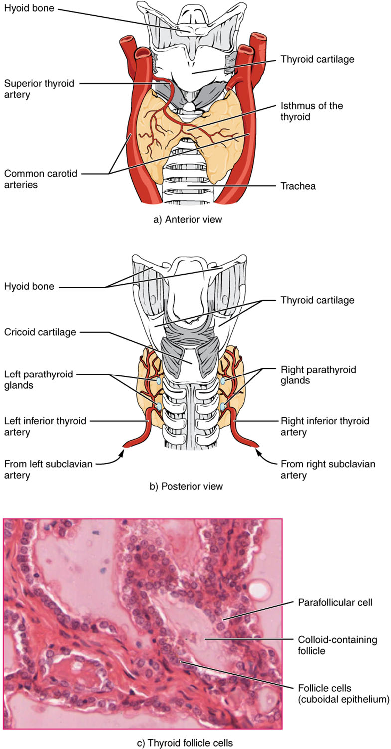
Hyoid bone The hyoid bone serves as an anchor point above the thyroid gland, providing structural support to the neck. It is a U-shaped bone that does not articulate directly with other bones but supports the tongue and larynx.
Thyroid cartilage The thyroid cartilage forms the prominent Adam’s apple, protecting the larynx and supporting the vocal cords. It also provides a framework that partially encases the upper portion of the thyroid gland.
Superior thyroid artery The superior thyroid artery supplies oxygenated blood to the upper part of the thyroid gland from the external carotid artery. This vessel is critical for delivering nutrients to support hormone synthesis.
Common carotid arteries The common carotid arteries run parallel to the thyroid gland, providing a major blood supply to the head and neck. They are essential for maintaining the gland’s metabolic activity.
Isthmus of the thyroid The isthmus of the thyroid is a narrow band of tissue connecting the gland’s two lobes across the trachea. This structure ensures the gland encircles the windpipe for efficient hormone distribution.
Trachea The trachea, or windpipe, lies centrally beneath the thyroid gland, facilitating airflow to the lungs. Its proximity to the thyroid allows for close interaction with surrounding structures.
Cricoid cartilage The cricoid cartilage forms the lower part of the larynx, sitting just below the thyroid cartilage. It provides a stable base for the thyroid gland and supports the airway.
Left parathyroid glands The left parathyroid glands are small endocrine glands embedded on the thyroid’s posterior surface, regulating calcium levels. They work in tandem with the thyroid to maintain mineral homeostasis.
Right parathyroid glands The right parathyroid glands mirror their left counterparts, secreting parathyroid hormone to control blood calcium. Their location ensures balanced hormonal regulation across the neck.
Left inferior thyroid artery The left inferior thyroid artery branches from the subclavian artery, supplying the lower thyroid lobe. It complements the superior artery in nourishing the gland.
Right inferior thyroid artery The right inferior thyroid artery provides blood to the lower right lobe, ensuring adequate circulation. Its origin from the subclavian artery supports thyroid function bilaterally.
From left subclavian artery The left subclavian artery is the source of the left inferior thyroid artery, delivering blood from the heart. This connection underscores the gland’s extensive vascular network.
From right subclavian artery The right subclavian artery supplies the right inferior thyroid artery, maintaining symmetry in blood flow. This dual supply enhances the thyroid’s resilience.
Parafollicular cell Parafollicular cells, or C cells, are located within the thyroid follicles and secrete calcitonin to lower blood calcium levels. These cells play a key role in calcium homeostasis alongside the parathyroids.
Colloid-containing follicle The colloid-containing follicle stores thyroid hormones T3 and T4 within a gel-like substance. This reservoir allows for controlled hormone release into the bloodstream.
Follicle cells (cuboidal epithelium) Follicle cells form the lining of thyroid follicles, synthesizing and secreting T3 and T4. Their cuboidal shape optimizes hormone production and storage.
Anatomical Overview of the Thyroid Gland
The thyroid gland’s location and structure are fundamental to its endocrine role. Positioned anterior to the trachea, it adapts to the neck’s anatomy for efficient function.
- The gland consists of two lobes connected by the isthmus, creating a butterfly-like shape.
- Blood supply from the superior and inferior thyroid arteries ensures robust hormonal output.
- The hyoid and thyroid cartilages provide structural support, protecting the gland from external pressure.
- The trachea’s central position allows the thyroid to influence respiratory and metabolic coordination.
Anterior and Posterior Perspectives
The anterior view highlights the thyroid’s external landmarks. This perspective reveals its relationship with the carotid arteries and superior thyroid artery.
- The thyroid cartilage and hyoid bone frame the gland’s upper boundary.
- The isthmus is visible, linking the lobes across the trachea.
- The posterior view exposes the parathyroid glands and inferior arteries.
- Cricoid cartilage supports the lower thyroid structure, enhancing stability.
Microscopic Structure of Thyroid Follicles
Thyroid follicles are the functional units of the gland at a cellular level. These structures are critical for hormone storage and release.
- Parafollicular cells regulate calcium by secreting calcitonin, complementing parathyroid activity.
- Colloid-containing follicles hold thyroglobulin, the precursor to T3 and T4.
- Follicle cells, with their cuboidal epithelium, actively produce thyroid hormones.
- The microscopic view at 1332x magnification reveals the intricate cellular matrix.
Physiological Role of the Thyroid Gland
The thyroid gland’s hormones, T3 and T4, regulate metabolism across the body. This function influences energy production, growth, and development.
- T3 (triiodothyronine) enhances cellular oxygen consumption, boosting metabolic rate.
- T4 (thyroxine) serves as a prohormone, converting to T3 in peripheral tissues.
- Calcitonin from parafollicular cells lowers blood calcium, preventing hypercalcemia.
- Hormonal imbalances can lead to conditions like hypothyroidism or hyperthyroidism.
Clinical Significance and Imaging
Understanding thyroid anatomy aids in diagnosing related disorders. Imaging techniques like ultrasound visualize gland size and nodules.
- The parathyroid glands’ proximity requires careful surgical consideration.
- Blood flow via carotid and subclavian arteries is assessed in vascular studies.
- Follicle cell activity is monitored through hormone level tests.
- Structural changes may indicate goiter or thyroid cancer, necessitating biopsy.
The thyroid gland’s intricate anatomy and cellular makeup underscore its critical role in maintaining bodily equilibrium. Its strategic location and robust vascular supply enable it to efficiently produce and distribute hormones, impacting overall health. Exploring these aspects provides valuable insights into its function and potential clinical interventions.

