Peristalsis is the fundamental mechanism by which food is propelled through our digestive system, a series of involuntary muscle contractions that ensure efficient nutrient processing. This article will explore the mechanics of peristalsis, detailing how alternating waves of muscle contraction and relaxation facilitate the unidirectional movement of food, highlighting its critical role in digestion and overall gastrointestinal health.
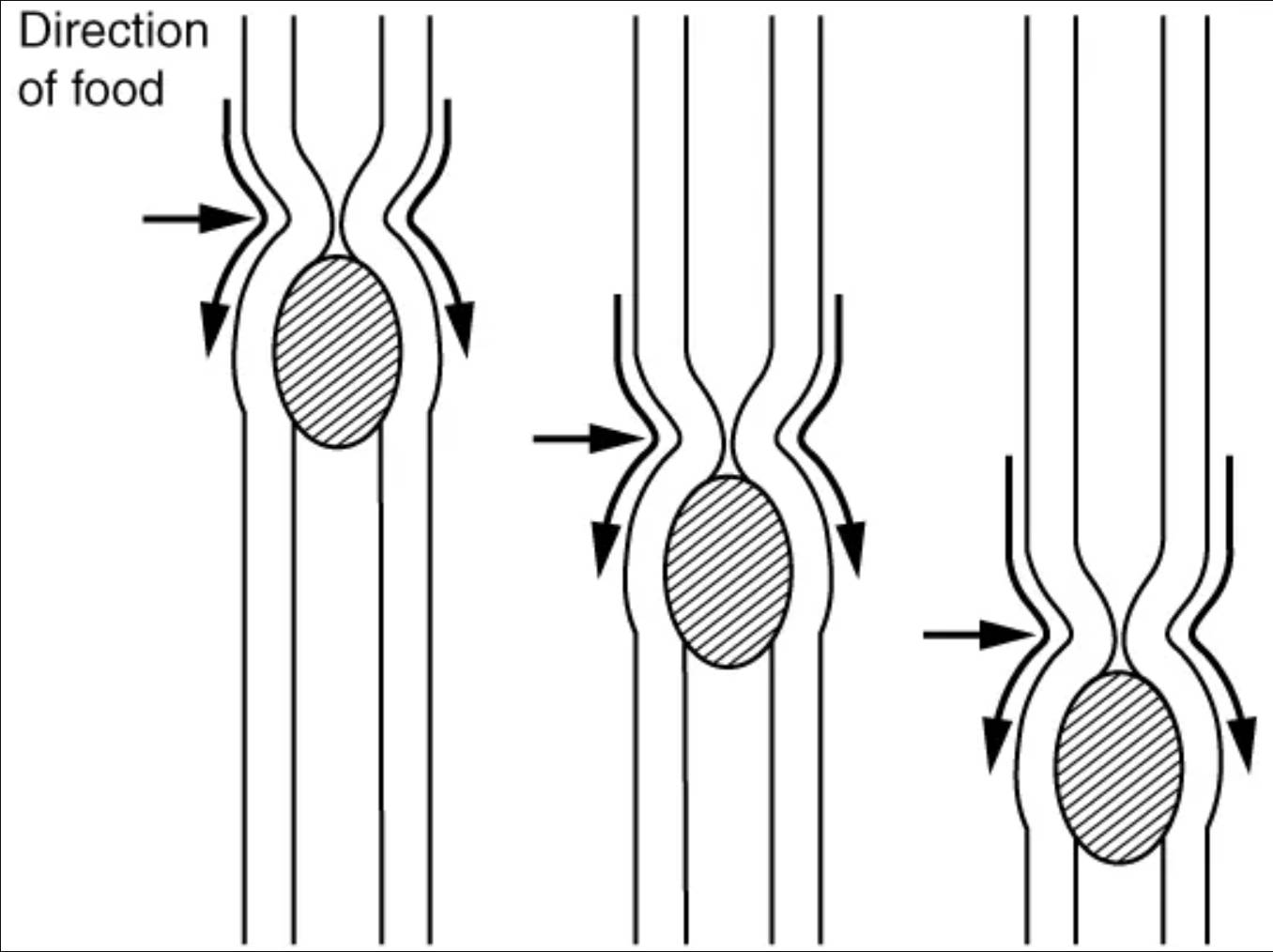
Direction of food: This label indicates the overall path that the food bolus takes as it moves through the digestive tract. In peristalsis, food moves in a unidirectional manner, typically from the mouth towards the anus, driven by coordinated muscle actions. This ensures efficient processing and absorption of nutrients throughout the alimentary canal.
The image vividly demonstrates the process of peristalsis, showing a bolus of food moving through a tubular structure. The arrows around the bolus illustrate the forces at play:
- Top arrows: These indicate the contraction of muscles behind the food bolus, pushing it forward.
- Bottom arrows: These show the relaxation of muscles in front of the bolus, allowing it to move into the widened segment.
This sequential contraction and relaxation is the hallmark of peristaltic movement, ensuring that food progresses steadily and efficiently through the digestive system. The rhythmic nature of these muscle actions prevents reflux and optimizes the time available for digestion and absorption.
The human digestive system is a marvel of biological engineering, designed to efficiently process the food we consume and extract vital nutrients. A cornerstone of this efficiency is peristalsis, an involuntary, wave-like muscular contraction that propels food through the alimentary canal. This sophisticated mechanism ensures that once food is swallowed, it continues its journey through the esophagus, stomach, and intestines, regardless of body position. Without peristalsis, food would remain static, leading to severe digestive dysfunction and impaired nutrient absorption.
Peristalsis is not a simple squeezing action but a highly coordinated sequence of events orchestrated by the smooth muscles of the digestive tract and regulated by the enteric nervous system. It involves two primary types of muscle layers:
- Circular muscles: These muscles contract behind the food bolus, narrowing the lumen and pushing the food forward.
- Longitudinal muscles: These muscles contract in front of the food bolus, shortening the segment and widening the lumen to receive the food.
This precise interplay of contraction and relaxation creates a propulsion wave, ensuring unidirectional movement and preventing the backward flow of digested material.
The Mechanics of Peristalsis: A Coordinated Movement
The fundamental principle of peristalsis is the alternating contraction and relaxation of the smooth muscle layers that constitute the wall of the digestive tract. When a bolus of food enters a section of the alimentary canal, the circular muscles immediately behind the bolus contract. This contraction applies pressure to the food, effectively squeezing it forward. Simultaneously, the longitudinal muscles in front of the bolus contract, causing that segment of the tube to shorten and widen. This widening action creates a low-pressure zone, making it easier for the food bolus to advance. As the food moves, the wave of contraction and relaxation follows it, ensuring a continuous and unidirectional progression. This rhythmic process is incredibly efficient, allowing food to traverse the entire digestive tract, from the esophagus to the large intestine, with precision.
Neural Control and Regulation of Peristalsis
The intricate coordination of peristalsis is primarily governed by the enteric nervous system (ENS), often referred to as the “second brain” of the gut. This intrinsic nervous system operates largely independently, though it can be modulated by the central nervous system. Within the ENS, two major plexuses are crucial: the myenteric plexus (Auerbach’s plexus), located between the circular and longitudinal muscle layers, and the submucosal plexus (Meissner’s plexus). The myenteric plexus is primarily responsible for controlling the strength and frequency of muscle contractions, directly regulating the propulsive movements of peristalsis. Afferent neurons detect the presence and distension caused by the food bolus, triggering efferent neurons to stimulate the appropriate muscle responses. This complex neural network ensures that peristalsis is adaptable, adjusting to the volume and consistency of the ingested food.
Clinical Significance: When Peristalsis Goes Awry
Dysfunction in peristalsis can lead to a variety of gastrointestinal disorders, highlighting the critical importance of this physiological process. For instance, achalasia is a condition where the lower esophageal sphincter fails to relax, and the esophagus loses its normal peristaltic ability, making it difficult for food to pass into the stomach. This can lead to dysphagia (difficulty swallowing), regurgitation, and chest pain. Similarly, conditions like gastroparesis, often seen in individuals with diabetes, involve delayed gastric emptying due to impaired peristaltic contractions in the stomach. Irritable bowel syndrome (IBS) can also involve abnormal peristaltic patterns, leading to symptoms like diarrhea, constipation, and abdominal pain. Understanding the mechanisms of peristalsis is therefore crucial for diagnosing and managing these and other digestive motility disorders, ultimately improving patient outcomes.
In conclusion, peristalsis is a fundamental, involuntary motor activity of the digestive system, essential for the efficient movement of food and the subsequent absorption of nutrients. Through the coordinated action of circular and longitudinal smooth muscles, meticulously regulated by the enteric nervous system, food is rhythmically propelled along the alimentary canal. Any disruption to this vital process can have significant health implications, underscoring the importance of healthy gastrointestinal motility. The continuous, wave-like nature of peristalsis ensures that our digestive system functions optimally, supporting overall health and well-being.

