Unveiling the Rattlesnake’s Spine: A Look at Procoelous Vertebrae
Delve into the specialized anatomy of a rattlesnake’s procoelous vertebrae, a key adaptation that grants these reptiles their exceptional flexibility and strength. This article explores the unique structural features, such as the convex protrusion and concave socket, that enable the snake’s characteristic movement. Understanding these intricate details provides insight into the evolutionary marvel of serpentine locomotion and skeletal design.
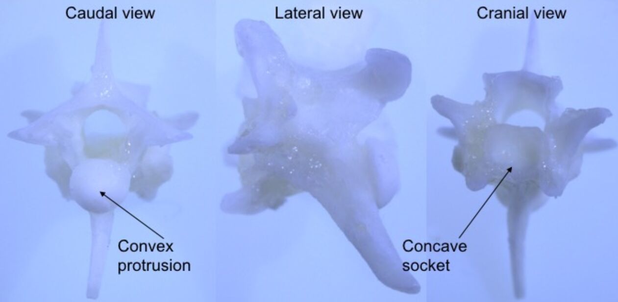
Convex protrusion: This refers to the rounded, anterior projection found on the caudal (posterior) end of a procoelous vertebra. This convex surface articulates perfectly with the concave socket of the subsequent vertebra, forming a highly mobile ball-and-socket joint. This anatomical feature is crucial for the snake’s remarkable flexibility, allowing for complex undulatory movements.
Concave socket: Located on the cranial (anterior) end of a procoelous vertebra, this indentation receives the convex protrusion of the preceding vertebra. The deep curvature of the socket ensures a secure yet fluid articulation, facilitating a wide range of motion without sacrificing stability. This unique joint design is fundamental to the rattlesnake’s ability to coil, strike, and navigate diverse terrains.
The skeletal system of snakes, particularly their vertebral column, is a testament to evolutionary adaptation, enabling their unique limbless locomotion. Unlike mammals or birds, snakes possess an exceptionally long and flexible spine composed of hundreds of individual vertebrae. These vertebrae, especially those in the trunk region, are predominantly procoelous, a term describing their specific articulating surfaces. This specialized structure is pivotal for the snake’s ability to undulate, constrict, and propel itself across various environments with remarkable efficiency and power.
Procoelous vertebrae are characterized by a concave anterior (cranial) end and a convex posterior (caudal) end. This specific configuration creates a ball-and-socket type of joint between adjacent vertebrae, allowing for extensive movement in multiple planes—dorsoventral, lateral, and rotational. Such a design ensures maximum flexibility without compromising the structural integrity of the spinal column. The robust nature of these articulations also provides significant resistance to dislocation, a crucial factor for an animal that relies entirely on its body for movement and subduing prey.
The intricate arrangement of these vertebrae, coupled with powerful musculature, allows snakes to perform complex movements. Whether it’s the sinuous gliding of sidewinding, the powerful constriction used to subdue prey, or the rapid strike of a predator like the rattlesnake, the procoelous vertebral column is at the heart of these actions. Each vertebra, while individually small, contributes collectively to the immense flexibility and strength that defines serpentine locomotion, making them highly effective predators and survivors in diverse ecosystems.
Key features of rattlesnake procoelous vertebrae include:
- Concave anterior surface: Facilitates articulation.
- Convex posterior surface: Forms a ball-and-socket joint.
- Neural arch: Protects the spinal cord.
- Transverse processes: Attachment points for ribs and muscles.
- Zygapophyses: Interlocking processes providing stability.
In conclusion, the procoelous vertebrae of the rattlesnake exemplify a pinnacle of evolutionary engineering, perfectly adapted for a life of limbless locomotion. The precise articulation of the convex protrusion into the concave socket provides both the extraordinary flexibility and robust stability necessary for a snake to navigate complex terrains, launch rapid strikes, and exert powerful constricting forces. This intricate skeletal design underscores the unique anatomical specializations that enable snakes to thrive as formidable predators and demonstrate a fascinating departure from the typical vertebrate body plan.

