The human throat and nasal cavity form a vital part of the respiratory and digestive systems, with structures like the tonsils playing a key role in immune defense. This article provides a detailed examination of a lateral sectional view of the throat, highlighting anatomical features such as the nasal cavity, tonsils, and pharyngeal regions, alongside insights into the gross pathology of hypertrophic tonsils. Medical professionals, students, and curious individuals can gain a deeper understanding of these structures’ functions and clinical relevance through the accompanying diagram and pathology images.
Anatomical Structures of the Throat and Nasal Cavity: Labeled Diagram Explanation
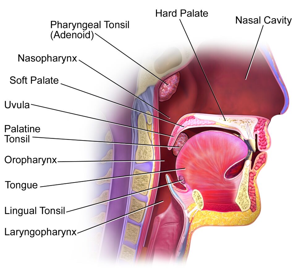
The diagram illustrates a lateral sectional view of the throat and nasal cavity, identifying key anatomical components with specific roles. Below is a detailed explanation of each labeled part.
Nasal Cavity
The nasal cavity is the upper respiratory passageway located above the hard palate, filtering, warming, and humidifying inhaled air before it reaches the lungs. It also houses olfactory receptors responsible for the sense of smell, contributing to taste perception through neural connections.
Hard Palate
The hard palate forms the rigid anterior roof of the mouth, separating the oral cavity from the nasal cavity and providing structural support for chewing. It is composed of bone covered by a mucous membrane, aiding in the initial stages of swallowing by elevating to direct food backward.
Pharyngeal Tonsil (Adenoid)
The pharyngeal tonsil, commonly known as the adenoid, is a lymphoid tissue located in the nasopharynx, serving as a first line of defense against inhaled pathogens. Enlargement, or adenoid hypertrophy, can obstruct nasal breathing and lead to conditions like chronic sinusitis or sleep-disordered breathing.
Nasopharynx
The nasopharynx is the upper portion of the pharynx behind the nasal cavity, connecting to the nasal passages and facilitating air passage to the lower respiratory tract. It contains the adenoids and the opening of the Eustachian tubes, which regulate middle ear pressure, making it prone to infections like nasopharyngitis.
Soft Palate
The soft palate is a flexible muscular structure at the back of the roof of the mouth, closing off the nasopharynx during swallowing to prevent nasal regurgitation. It also plays a role in speech by controlling airflow, with dysfunction potentially causing hypernasal speech in conditions like cleft palate.
Uvula
The uvula is a small, pendulous projection from the soft palate, assisting in speech articulation by modulating airflow and preventing food from entering the nasal cavity during swallowing. Its movement triggers the gag reflex, a protective mechanism to expel foreign objects from the throat.
Palatine Tonsil
The palatine tonsils are lymphoid tissues on either side of the oropharynx, part of the Waldeyer’s ring, trapping pathogens to initiate immune responses. They are commonly affected by infections like tonsillitis, which can lead to hypertrophy and require medical or surgical intervention.
Oropharynx
The oropharynx is the middle pharyngeal region behind the oral cavity, serving as a passageway for both air and food. It contains the palatine and lingual tonsils, making it a critical area for immune surveillance and prone to conditions like pharyngitis.
Tongue
The tongue is a muscular organ at the floor of the mouth, essential for taste, speech, and swallowing by manipulating food into a bolus. Its innervation by the hypoglossal nerve (cranial nerve XII) enables precise movements, with taste buds on its surface detecting sweet, sour, salty, bitter, and umami flavors.
Lingual Tonsil
The lingual tonsil is lymphoid tissue located at the base of the tongue, contributing to local immune defense against ingested pathogens. Its enlargement can cause discomfort or difficulty swallowing, occasionally necessitating diagnostic evaluation for underlying conditions.
Laryngopharynx
The laryngopharynx is the lower part of the pharynx, connecting to the larynx and esophagus, directing air to the lungs and food to the digestive tract. Its strategic location makes it susceptible to reflux esophagitis or laryngopharyngeal reflux, impacting voice quality and swallowing.
Gross Pathology of Hypertrophic Tonsils: A Detailed Analysis
The pathology images offer a close look at a fresh hypertrophic tonsil, revealing its surfaces and internal composition. This section provides a professional insight into its clinical and anatomical features.
- Surface Facing the Aerodigestive Tract (Top Left): This exposed surface features crypts that can harbor bacteria and debris, often leading to inflammation or infection in hypertrophic tonsils. Obstruction from enlargement may result in sleep apnea or dysphagia, necessitating intervention.
- Opposite Surface (Top Right, Cauterized): The cauterized surface suggests surgical treatment, likely to reduce tonsil size or control bleeding during tonsillectomy. This technique seals tissue, minimizing postoperative hemorrhage, a common concern in tonsil surgery.
- Cut Sections (Bottom): Internal sections display hyperplastic lymphoid follicles, indicative of chronic immune activation in hypertrophic tonsils. These are examined histopathologically to exclude malignancies like non-Hodgkin lymphoma in persistent cases.
Anatomical Introduction: Throat and Nasal Cavity Functions
The throat and nasal cavity structures are integral to respiratory, digestive, and immune functions, working in harmony to maintain health. This section explores their roles and physical characteristics in a clinical context.
- Respiratory Role of the Nasal Cavity and Pharynx: The nasal cavity filters air through mucous membranes and cilia, while the nasopharynx and laryngopharynx ensure smooth airflow to the lungs. Any obstruction, such as adenoid hypertrophy, can impair breathing and oxygen saturation.
- Digestive Pathway Support: The oropharynx and laryngopharynx facilitate food passage, with the tongue and soft palate coordinating swallowing to prevent aspiration. Dysfunction, as seen in neurological disorders, can lead to choking or malnutrition.
- Immune Defense Mechanism: Tonsils, including the pharyngeal, palatine, and lingual types, form the Waldeyer’s ring, producing lymphocytes to combat pathogens. Their hypertrophy often signals chronic exposure to infection, requiring monitoring.
- Physical Characteristics: The hard palate’s bony structure contrasts with the soft palate’s muscular flexibility, while the uvula’s small size belies its role in speech and reflex action. The tongue’s mobility and tonsillar lymphoid tissue highlight their dynamic functions.
Clinical Relevance of Hypertrophic Tonsils
Hypertrophic tonsils, as shown in the pathology images, are a significant condition affecting throat function and overall health. This section outlines its implications and management for medical professionals.
- Causes and Risk Factors: Chronic infections from Streptococcus or viruses like Epstein-Barr, along with allergies or genetic predisposition, drive tonsil hypertrophy. Environmental factors, such as polluted air, may exacerbate lymphoid tissue enlargement.
- Symptoms and Complications: Patients may experience snoring, obstructive sleep apnea, or recurrent tonsillitis, with severe cases linked to cardiovascular strain from hypoxia. Persistent enlargement warrants investigation for malignancy.
- Diagnostic Techniques: Clinical grading (e.g., Brodsky scale) assesses tonsil size, while endoscopy or imaging evaluates airway obstruction. Biopsies of cut sections help rule out lymphoma or other pathologies.
- Treatment Strategies: Antibiotics manage acute infections, while tonsillectomy is indicated for chronic hypertrophy or obstruction. Postoperative care focuses on pain control and monitoring for bleeding, typically resolving within two weeks.
Surgical and Therapeutic Insights for Tonsil Management
Tonsillectomy is a standard procedure for hypertrophic tonsils, offering relief from obstruction and infection. This section details the process and considerations for healthcare providers.
- Indications for Surgery: Recurrent tonsillitis (over 6 episodes annually) or severe sleep apnea justifies tonsillectomy. Peritonsillar abscess unresponsive to drainage also prompts surgical evaluation.
- Surgical Methods: Electrocautery, as seen in the cauterized surface, reduces bleeding, while cold knife dissection preserves tissue integrity. Coblation uses radiofrequency for less thermal damage, enhancing recovery.
- Postoperative Care: Patients follow a soft diet and hydration regimen, with acetaminophen for pain. Bleeding risk peaks within 10 days, requiring vigilant follow-up.
- Long-Term Outcomes: Surgery often resolves sleep apnea and improves quality of life, though rare regrowth of lingual tonsils may occur, necessitating further assessment.
The lateral sectional view of the throat and nasal cavity, combined with insights into hypertrophic tonsil pathology, underscores the intricate balance of these structures in health and disease. Medical professionals can leverage this knowledge to diagnose and treat related conditions effectively, enhancing patient care and outcomes on May 11, 2025, and beyond.
- Nasal Cavity and Throat Anatomy: A Lateral View Guide
- Hypertrophic Tonsils Pathology: Understanding Throat Structures
- Tonsils and Pharynx: Anatomical Insights and Clinical Relevance
- Exploring Throat Anatomy: Nasal Cavity to Laryngopharynx
- Hypertrophic Tonsils: Anatomy, Pathology, and Treatment Options

