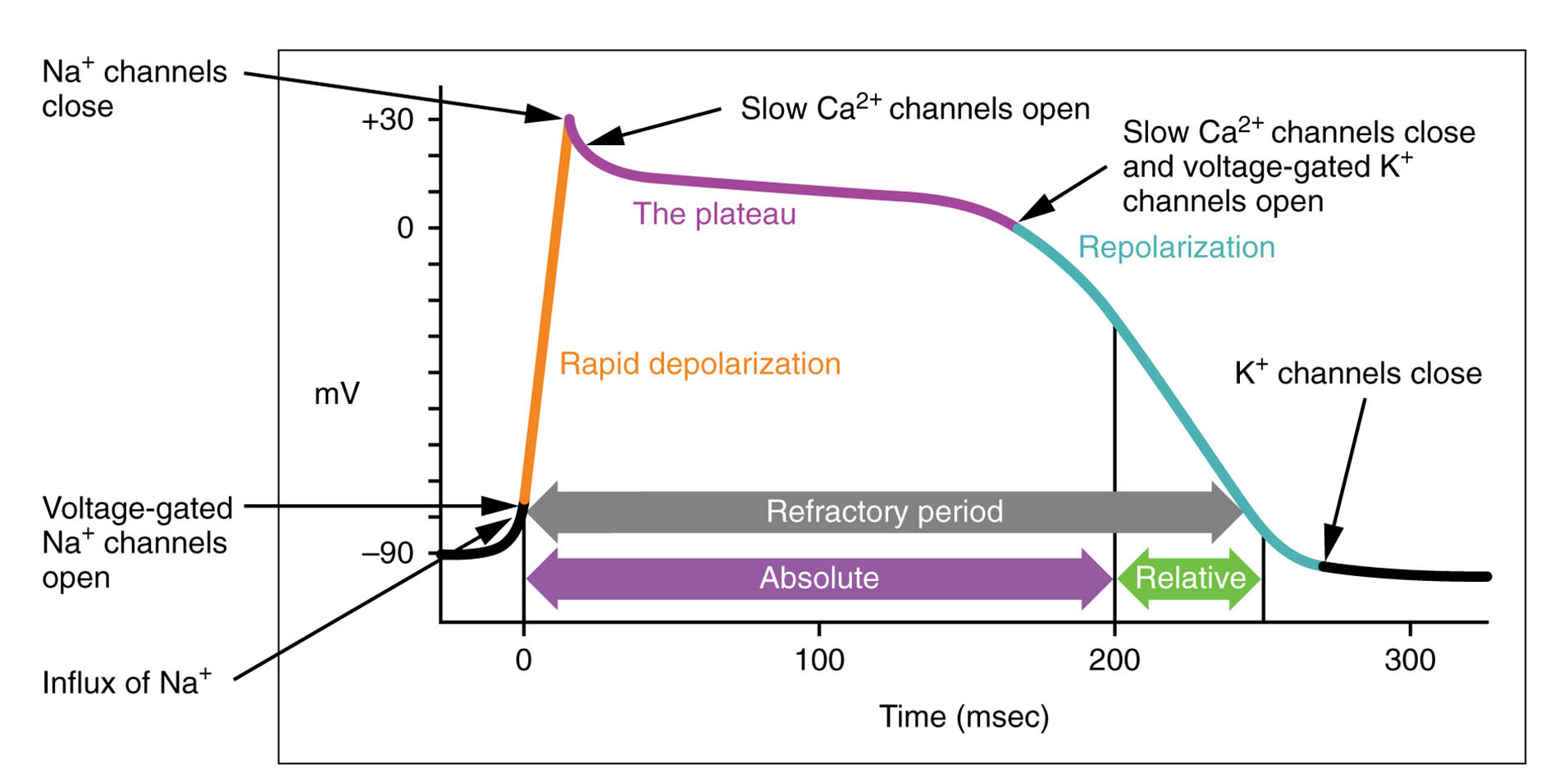
The action potential in cardiac cells is a fascinating process that underpins the heart’s rhythmic contractions, with a distinctive long plateau phase driven by calcium ion influx. This diagram highlights the long plateau phase and extended refractory period, illustrating how these features ensure the heart completes its contraction cycle effectively. Exploring this image provides a deeper understanding of the electrophysiological mechanisms that sustain cardiac function.
Labelled Parts Explanation
- Long plateau phase The long plateau phase is a prolonged depolarization period caused by a significant influx of calcium ions through L-type calcium channels. It sustains the contractile cells’ membrane potential, allowing the heart muscle to maintain tension during systole.
- Extended refractory period The extended refractory period follows the plateau phase, preventing another action potential until the cell fully relaxes due to a long absolute refractory period. This ensures the heart completes its contraction and refilling before the next beat, avoiding tetanic contraction.
Anatomical Overview of the Cardiac Action Potential
The cardiac action potential is uniquely designed to support the heart’s continuous pumping action. This diagram focuses on the key phases that distinguish cardiac cells from other muscle types.
- The long plateau phase maintains muscle contraction, driven by calcium influx.
- The extended refractory period protects the heart from premature re-excitation.
- These features are integral to the cardiac cycle’s efficiency.
- The process reflects the heart’s adaptation to its relentless workload.
This structure ensures effective blood circulation.
Role of the Long Plateau Phase
The long plateau phase is critical for cardiac contraction. Its duration supports the heart’s pumping action.
- The long plateau phase results from calcium entering the cell, triggering further release from the sarcoplasmic reticulum.
- This prolonged depolarization keeps the muscle contracted during ventricular ejection.
- The phase lasts 200-300 ms, much longer than in skeletal muscle.
- Calcium’s role enhances the force of contraction.
This mechanism is essential for sustaining circulation.
Significance of the Extended Refractory Period
The extended refractory period prevents overstimulation of the heart. This feature ensures proper rhythm.
- The extended refractory period aligns with the mechanical contraction, lasting nearly as long as systole.
- It includes a long absolute refractory period, blocking additional action potentials.
- This prevents tetanus, which would halt blood flow.
- The period allows complete ventricular relaxation before the next cycle.
This protection is vital for cardiac stability.
Ionic Basis of the Action Potential
The action potential in cardiac cells relies on specific ion movements. These shifts drive the electrical and contractile events.
- The long plateau phase is sustained by calcium influx through L-type channels.
- Rapid depolarization begins with sodium influx, initiating the action potential.
- The extended refractory period involves slow potassium efflux during repolarization.
- Calcium release amplifies the contractile force during the plateau.
This ionic interplay supports heart function.
Physiological Importance of the Plateau and Refractory Period
The plateau and refractory period optimize the heart’s pumping efficiency. Their design ensures continuous circulation.
- The long plateau phase provides time for ventricular ejection and atrial filling.
- The extended refractory period prevents arrhythmias by blocking re-excitation.
- These phases synchronize electrical and mechanical events.
- The heart adapts to varying demands through this mechanism.
This coordination is crucial for cardiovascular health.
Comparison with Other Muscle Types
The cardiac action potential differs from skeletal and smooth muscle. This distinction highlights its unique role.
- The long plateau phase is absent in skeletal muscle, which has a brief action potential.
- The extended refractory period ensures the heart avoids tetanus, unlike skeletal muscle.
- Cardiac cells rely on calcium for the plateau, while skeletal muscle depends on sodium.
- This design supports the heart’s rhythmic, involuntary contractions.
The difference reflects functional specialization.
Clinical Relevance of the Action Potential Phases
Understanding the action potential phases aids in managing cardiac conditions. These features are key clinical markers.
- Prolongation of the long plateau phase can lead to long QT syndrome, increasing arrhythmia risk.
- Shortening the extended refractory period may cause re-entrant tachycardias.
- Electrocardiograms monitor these phases for abnormalities.
- Treatments adjust ion channel activity to normalize the action potential.
This knowledge guides effective cardiac interventions.
Conclusion
The long plateau phase due to the influx of calcium ions – action potential in cardiac cells diagram offers a detailed view of the electrical processes that drive the heart’s contractions. By exploring the long plateau phase and extended refractory period, one gains insight into how these phases ensure the heart’s rhythmic efficiency and prevent overstimulation. This understanding serves as a foundation for studying cardiovascular physiology and addressing related health issues, encouraging further exploration of the heart’s intricate electrophysiological design and its critical role in sustaining life.

