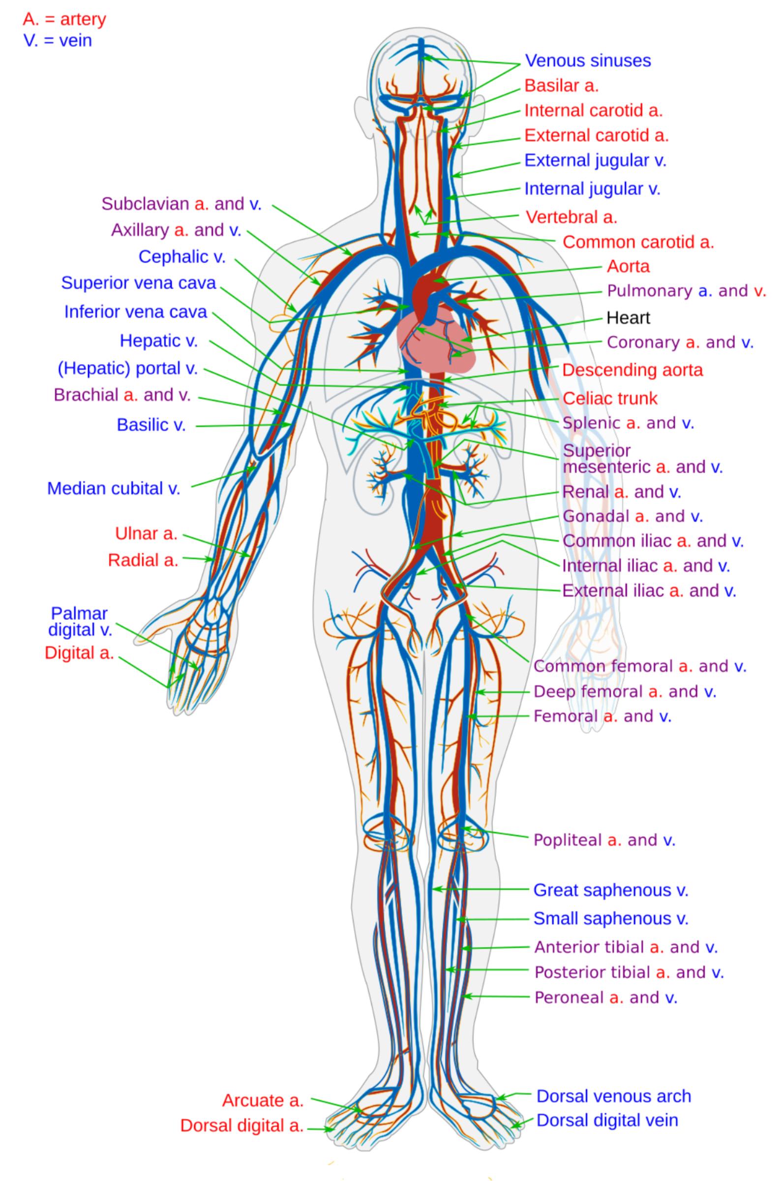
Explore the intricate network of the human circulatory system through this detailed diagram, highlighting major arteries (red) and veins (blue) throughout the body. Understand how this vital system transports oxygen, nutrients, hormones, and waste products, maintaining bodily functions and overall health. Delve into the anatomical pathways that ensure continuous blood flow from the heart to every cell and back again.
A.: artery: This abbreviation refers to an artery, a blood vessel that carries oxygenated blood away from the heart to the rest of the body. Arteries typically have thick, muscular walls to withstand the high pressure of blood being pumped.
V.: vein: This abbreviation denotes a vein, a blood vessel that carries deoxygenated blood back towards the heart. Veins have thinner walls and contain valves to prevent the backflow of blood, especially in the limbs.
Venous sinuses: These are specialized venous channels found primarily in the dura mater of the brain, collecting deoxygenated blood from the brain and draining into the internal jugular veins. Unlike other veins, they lack valves and typically don’t have muscular walls.
Basilar a.: This refers to the basilar artery, formed by the union of the two vertebral arteries at the base of the brain. It is a major blood vessel that supplies oxygenated blood to the brainstem, cerebellum, and posterior cerebral hemispheres.
Internal carotid a.: The internal carotid artery is a major paired artery in the head and neck, supplying blood to the brain, eyes, and other facial structures. It arises from the common carotid artery and does not give off branches in the neck.
External carotid a.: This artery is one of the two main branches of the common carotid artery, primarily supplying blood to the face, scalp, tongue, and other superficial structures of the head and neck. It has numerous branches that extend to these regions.
External jugular v.: The external jugular vein collects deoxygenated blood from the superficial parts of the head and neck. It drains into the subclavian vein, providing a pathway for venous return from these areas.
Internal jugular v.: The internal jugular vein is a large vein in the neck that collects deoxygenated blood from the brain, face, and neck. It runs alongside the common carotid artery and plays a crucial role in cranial venous drainage.
Vertebral a.: The vertebral artery is a major artery that arises from the subclavian artery and ascends through the neck vertebrae before entering the skull. It supplies blood to the brainstem, cerebellum, and posterior cerebrum, forming the basilar artery.
Common carotid a.: The common carotid artery is a major artery in the neck that bifurcates into the internal and external carotid arteries. It supplies blood to the head and neck and is a common site for pulse palpation.
Aorta: The largest artery in the body, the aorta originates from the left ventricle of the heart and arches superiorly before descending through the chest and abdomen. It distributes oxygenated blood to all systemic arteries.
Pulmonary a. and v.: The pulmonary artery carries deoxygenated blood from the right ventricle to the lungs, where it becomes oxygenated. The pulmonary veins then carry this newly oxygenated blood from the lungs back to the left atrium of the heart.
Heart: The central organ of the circulatory system, the heart is a muscular pump responsible for propelling blood throughout the body. It consists of four chambers that work in a coordinated rhythm to ensure continuous circulation.
Coronary a. and v.: The coronary arteries supply oxygenated blood directly to the heart muscle (myocardium) itself, while the coronary veins collect deoxygenated blood from the heart muscle and return it to the right atrium. These vessels are vital for the heart’s own nourishment.
Descending aorta: This is the portion of the aorta that extends from the aortic arch downwards through the chest (thoracic aorta) and abdomen (abdominal aorta). It gives off numerous branches to supply blood to the torso, abdomen, and lower limbs.
Subclavian a. and v.: The subclavian artery supplies blood to the arms, neck, and upper chest, while the subclavian vein drains blood from the same regions. These vessels pass under the clavicle (collarbone) and are crucial for upper limb circulation.
Axillary a. and v.: The axillary artery is a continuation of the subclavian artery in the axilla (armpit), supplying blood to the shoulder, chest wall, and arm. The axillary vein drains blood from these areas and becomes the subclavian vein.
Cephalic v.: The cephalic vein is a large, superficial vein that runs along the lateral side of the upper limb, from the hand to the shoulder. It is often visible through the skin and is a common site for venipuncture.
Superior vena cava: This large vein carries deoxygenated blood from the upper half of the body (head, neck, and arms) back to the right atrium of the heart. It is one of the two main venae cavae.
Inferior vena cava: The largest vein in the body, the inferior vena cava carries deoxygenated blood from the lower half of the body (torso, abdomen, and lower limbs) back to the right atrium. It ascends through the abdomen and diaphragm.
Hepatic v.: The hepatic veins are responsible for draining deoxygenated blood from the liver into the inferior vena cava. These veins are crucial for the liver’s role in processing nutrients and detoxifying blood.
(Hepatic) portal v.: The hepatic portal vein carries nutrient-rich, deoxygenated blood from the gastrointestinal tract, spleen, and pancreas to the liver. This unique system allows the liver to process absorbed nutrients and filter toxins before blood enters the general circulation.
Brachial a. and v.: The brachial artery is the main artery of the upper arm, supplying blood to the forearm and hand. The brachial veins typically accompany the artery, draining blood from the same regions.
Basilic v.: The basilic vein is a large, superficial vein that runs along the medial side of the upper limb. It typically joins with the brachial veins to form the axillary vein, contributing to venous return from the arm.
Median cubital v.: This vein connects the cephalic and basilic veins in the antecubital fossa (inner elbow region). It is a very common site for drawing blood and administering intravenous fluids due to its superficial location and size.
Ulnar a.: The ulnar artery is one of the two main arteries of the forearm, running along the medial side. It supplies blood to the medial forearm and contributes to the arterial supply of the hand, forming part of the superficial palmar arch.
Radial a.: The radial artery is the other main artery of the forearm, running along the lateral side. It is commonly used to take a pulse and supplies blood to the lateral forearm and hand, forming part of the deep palmar arch.
Palmar digital v.: These small veins drain deoxygenated blood from the fingers into the larger veins of the hand. They are part of the intricate venous network in the hand.
Digital a.: The digital arteries are small arteries that supply oxygenated blood to the fingers and toes. They branch off from larger arteries in the hand and foot.
Celiac trunk: This is a major arterial branch of the abdominal aorta, typically the first unpaired branch. It quickly divides into three main arteries: the splenic artery, the left gastric artery, and the common hepatic artery, supplying blood to the stomach, spleen, liver, and parts of the pancreas and duodenum.
Splenic a. and v.: The splenic artery supplies oxygenated blood to the spleen, pancreas, and parts of the stomach. The splenic vein drains deoxygenated blood from these organs, eventually joining the superior mesenteric vein to form the hepatic portal vein.
Superior mesenteric a. and v.: The superior mesenteric artery supplies oxygenated blood to a large portion of the small intestine and parts of the large intestine. The superior mesenteric vein drains deoxygenated blood from these same regions and is a major tributary of the hepatic portal vein.
Renal a. and v.: The renal arteries supply oxygenated blood to the kidneys, while the renal veins drain deoxygenated blood from the kidneys into the inferior vena cava. These vessels are crucial for kidney function, including blood filtration and waste removal.
Gonadal a. and v.: Also known as testicular arteries/veins in males and ovarian arteries/veins in females, these vessels supply/drain blood from the gonads. The gonadal arteries branch directly from the aorta, and the veins drain into the inferior vena cava (right) or renal vein (left).
Common iliac a. and v.: The common iliac artery is formed by the bifurcation of the abdominal aorta and further divides into the internal and external iliac arteries. The common iliac vein is formed by the union of the internal and external iliac veins and drains into the inferior vena cava. These vessels are key for pelvic and lower limb circulation.
Internal iliac a. and v.: The internal iliac artery supplies blood to the pelvic organs, buttocks, and medial thigh. The internal iliac vein drains blood from these regions, contributing to the common iliac vein.
External iliac a. and v.: The external iliac artery continues as the femoral artery after passing the inguinal ligament, supplying blood to the lower limbs. The external iliac vein drains blood from the lower limb and becomes the common iliac vein.
Common femoral a. and v.: The common femoral artery is a continuation of the external iliac artery in the upper thigh, branching into the superficial and deep femoral arteries. The common femoral vein drains blood from the entire lower limb and becomes the external iliac vein.
Deep femoral a. and v.: Also known as the profunda femoris artery, this is a large branch of the common femoral artery, supplying blood to the deep muscles of the thigh. The deep femoral vein accompanies it, draining blood from these muscles.
Femoral a. and v.: The femoral artery is the main artery of the thigh, continuing from the external iliac artery. It supplies blood to the entire lower limb. The femoral vein accompanies the artery, serving as the primary deep vein for venous return from the leg and thigh.
Popliteal a. and v.: The popliteal artery is a continuation of the femoral artery behind the knee. It supplies blood to the knee joint and lower leg. The popliteal vein drains blood from these areas and becomes the femoral vein.
Great saphenous v.: This is the longest superficial vein in the body, running along the medial aspect of the entire lower limb, from the foot to the groin, where it drains into the femoral vein. It is often used for coronary artery bypass grafts.
Small saphenous v.: The small saphenous vein is a superficial vein that runs along the posterior aspect of the lower leg, draining blood from the lateral foot and ankle. It typically empties into the popliteal vein.
Anterior tibial a. and v.: The anterior tibial artery supplies blood to the anterior compartment of the lower leg and the top of the foot. The anterior tibial veins drain blood from these areas and join the posterior tibial veins to form the popliteal vein.
Posterior tibial a. and v.: The posterior tibial artery supplies blood to the posterior and lateral compartments of the lower leg and the sole of the foot. The posterior tibial veins drain blood from these regions.
Peroneal a. and v.: Also known as fibular artery and vein, these vessels supply and drain blood from the lateral compartment of the lower leg. They are branches of the posterior tibial vessels.
Arcuate a.: The arcuate artery is an artery in the foot, branching off the dorsalis pedis artery. It crosses the metatarsals and gives rise to dorsal metatarsal arteries, supplying blood to the toes.
Dorsal venous arch: This superficial venous network is located on the dorsum (top) of the foot. It collects blood from the digital veins and is the origin of the great and small saphenous veins.
Dorsal digital vein: These small veins drain deoxygenated blood from the toes into the dorsal venous arch of the foot. They are part of the intricate venous network in the foot.
The circulatory system is a complex and vital network responsible for transporting blood throughout the body. Comprising the heart, blood vessels (arteries, veins, and capillaries), and blood itself, this system ensures the delivery of oxygen and nutrients to tissues while simultaneously removing carbon dioxide and other metabolic waste products. The intricate web of arteries, depicted in red, carries oxygenated blood away from the heart, branching into smaller and smaller vessels to reach every cell. Conversely, veins, shown in blue, collect deoxygenated blood and waste products, returning them to the heart and lungs for reoxygenation.
This comprehensive diagram provides an anterior view of the major arterial and venous pathways in the human body. It illustrates the systemic circulation, which supplies blood to all tissues except the lungs, and parts of the pulmonary circulation, which involves blood flow to and from the lungs. Understanding the anatomical distribution of these vessels is crucial for medical professionals in diagnosing and treating various conditions, from cardiovascular diseases to peripheral vascular disorders. The precise naming and location of each vessel highlight the organized efficiency of this life-sustaining system.
The continuous flow of blood facilitated by this network is essential for maintaining homeostasis, regulating body temperature, and distributing hormones. Arteries, characterized by their strong, elastic walls, withstand the high pressure generated by the heart’s pumping action. Veins, with their thinner walls and presence of valves, operate under lower pressure and rely on muscle contractions and breathing to push blood back towards the heart. The detailed labeling in the image serves as a fundamental resource for learning and identifying key components of this indispensable biological system.
-
- The circulatory system transports oxygen, nutrients, and waste.
- Arteries carry oxygenated blood away from the heart.
- Veins return deoxygenated blood to the heart.
- The heart is the central pump of the system.
- This diagram illustrates major systemic vessels.
The Aorta: The Main Highway of Oxygenated Blood
The aorta stands as the largest artery in the human body, serving as the primary conduit for oxygenated blood leaving the left ventricle of the heart. Its journey begins with the ascending aorta, which gives rise to the coronary arteries that supply blood to the heart muscle itself. It then forms the aortic arch, from which three major branches emerge: the brachiocephalic trunk, the left common carotid artery, and the left subclavian artery, supplying the upper limbs, head, and neck. As it descends through the thorax (thoracic aorta) and abdomen (abdominal aorta), the aorta gives off numerous vital branches to supply all the major organs and lower extremities. For instance, the celiac trunk supplies the stomach, liver, and spleen, while the renal arteries supply the kidneys. The integrity of the aorta is paramount; conditions like aortic aneurysms, which involve a weakening and bulging of the aortic wall, can be life-threatening if they rupture, leading to massive internal bleeding.
The Vena Cavae: Major Return Routes for Deoxygenated Blood
Complementing the arterial system, the vena cavae represent the two largest veins in the body, responsible for collecting deoxygenated blood from the systemic circulation and returning it to the right atrium of the heart. The superior vena cava (SVC) gathers blood from the head, neck, upper limbs, and thorax. In contrast, the inferior vena cava (IVC) collects blood from the abdomen, pelvis, and lower limbs. These massive veins merge into the right atrium, completing the systemic circuit before the blood is pumped to the lungs for reoxygenation via the pulmonary artery. The functioning of these large veins is critical for maintaining venous return and preventing venous stasis, which can lead to conditions like deep vein thrombosis (DVT), where blood clots form, potentially leading to pulmonary embolism if they dislodge.
The intricate and expansive nature of the human circulatory system, with its specialized arterial and venous pathways, underscores its critical role in sustaining life. From the robust pumping action of the heart to the delicate exchange of gases and nutrients in capillaries, every component works in harmony. A comprehensive understanding of these anatomical structures and their physiological functions is indispensable for healthcare professionals, enabling accurate diagnosis, effective treatment, and the promotion of cardiovascular well-being.


![[1]](http://www.rci.rutgers.edu/%7Euzwiak/AnatPhys/Blood_Vessels_files/image040.jpg)
![[2]](http://a248.e.akamai.net/7/248/430/20061026230922/www.merck.com/pubs/mmanual_home2/cp/cp003.gif)
![[3]](http://www.trauma-med.net/Circulatorysystem.JPG)