The extraocular muscles are fundamental to eye movement and alignment, working seamlessly within the orbit to support vision. This anterior view of the right eye illustrates the six key muscles and their spatial arrangement, providing a window into their coordinated function.
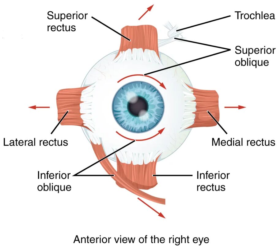
Superior rectus The superior rectus elevates the eye and assists in inward rotation, playing a key role in upward gaze. It is innervated by the oculomotor nerve, ensuring precise control during vertical movements.
Trochlea The trochlea is a pulley-like cartilage structure that guides the superior oblique tendon, enhancing its mechanical efficiency. It allows the superior oblique muscle to exert a unique downward and outward pull on the eye.
Superior oblique The superior oblique rotates the eye downward and outward, aiding in intorsion and downward gaze. Its tendon passes through the trochlea, enabling complex rotational movements.
Lateral rectus The lateral rectus abducts the eye, moving it outward away from the nose. Controlled by the abducens nerve, it is essential for lateral eye movements and peripheral vision.
Inferior oblique The inferior oblique elevates and outwardly rotates the eye, supporting upward and lateral gaze. Its insertion pattern allows for versatile tracking of objects in various directions.
Medial rectus The medial rectus adducts the eye, moving it inward toward the nose. Innervated by the oculomotor nerve, it balances the lateral rectus for horizontal alignment.
Inferior rectus The inferior rectus depresses the eye and aids in outward rotation, facilitating downward gaze. It works in opposition to the superior rectus for vertical eye positioning.
Anatomy of the Extraocular Muscles
The extraocular muscles form a sophisticated network around the eyeball, depicted here in an anterior view of the right eye. Their strategic placement ensures comprehensive control over eye movement and stability.
- The rectus muscles—superior, inferior, lateral, and medial—originate from the common tendinous ring within the orbit.
- The oblique muscles—superior and inferior—add torsional and diagonal movements, enhancing flexibility.
- The trochlea serves as a critical anchor for the superior oblique tendon, redirecting its force.
- Each muscle inserts into the sclera, the tough outer layer of the eyeball, at specific points.
- The orbit, a bony cavity, houses these muscles, providing protection and a range of motion.
- Blood supply from the ophthalmic artery nourishes these muscles, supporting their continuous activity.
- Connective tissue sheaths surround the muscles, reducing friction during movement.
Functions of Eye Movement
The extraocular muscles enable a wide range of eye movements, essential for vision and spatial orientation. Their coordinated action ensures smooth tracking and stable focus.
- The superior rectus lifts the eye, aiding in looking up or focusing on elevated objects.
- The lateral rectus moves the eye outward, crucial for following moving targets or scanning peripherally.
- The inferior rectus lowers the eye, supporting downward gaze like reading or inspecting the ground.
- The medial rectus brings the eye inward, essential for close-up vision and convergence.
- The superior oblique, via the trochlea, rotates the eye downward and outward for fine adjustments.
- The inferior oblique elevates and rotates the eye, facilitating upward and lateral tracking.
- These muscles work in pairs, with agonists and antagonists maintaining balance and alignment.
- Rapid saccadic movements rely on this muscular synergy for quick shifts in visual attention.
Role of the Trochlea in Muscle Function
The trochlea plays a pivotal role in directing the superior oblique muscle’s action. Its pulley mechanism enhances the muscle’s ability to control eye rotation.
- The trochlea redirects the superior oblique tendon, optimizing its pull angle for effective movement.
- This structure is located on the medial wall of the orbit, made of cartilage for durability.
- The tendon slides through the trochlea, reducing wear and ensuring smooth motion.
- Damage to the trochlea can impair superior oblique function, affecting downward gaze.
- The pulley system allows for a mechanical advantage, amplifying the muscle’s torque.
- Its position remains stable, providing a consistent anchor point for tendon action.
Innervation and Neural Control
The extraocular muscles are innervated by cranial nerves, ensuring precise and rapid responses to visual stimuli. This neural integration supports seamless eye coordination.
- The oculomotor nerve (CN III) supplies the superior, inferior, and medial rectus muscles.
- The trochlear nerve (CN IV) innervates the superior oblique, guiding its unique path through the trochlea.
- The abducens nerve (CN VI) controls the lateral rectus, specializing in abduction.
- These nerves originate from the brainstem, with nuclei coordinating bilateral eye movements.
- Reflexes like the vestibulo-ocular reflex stabilize gaze during head motion using these muscles.
- Nerve damage can lead to strabismus or diplopia, though this image depicts normal anatomy.
Clinical Relevance of Extraocular Muscles
Understanding the anterior view of the extraocular muscles aids in diagnosing and managing eye movement disorders. This image showcases healthy anatomy, serving as a reference for clinical practice.
- Weakness in the lateral rectus can cause esotropia, where the eye turns inward excessively.
- Superior oblique dysfunction, linked to trochlea issues, may result in vertical diplopia or cyclotorsion.
- The inferior rectus’s impairment can hinder downward gaze, impacting daily activities.
- Muscle imbalance is assessed with tests like the cover-uncover test or eye movement tracking.
- Surgical interventions, such as muscle recession, correct alignment in strabismus cases.
- Rehabilitation exercises strengthen these muscles after nerve palsy or injury.
- Electromyography evaluates muscle and nerve function for precise diagnosis.
In conclusion, the extraocular muscles, as illustrated in this anterior view, form a remarkable system for eye movement and stability. Their intricate design, supported by structures like the trochlea and precise innervation, underscores the complexity of maintaining clear vision and spatial awareness.

