The cerebellum, a key component of the hindbrain, plays an essential role in coordinating movement, balance, and motor learning. This image presents a detailed anatomical illustration of the cerebellum alongside an imaging view, highlighting structures such as the cerebellum, pons, inferior olive, and deep cerebellar white matter (arbor vitae), which facilitate sensory input and output pathways. Exploring these elements offers a comprehensive understanding of cerebellar function and its integration with the brain stem.
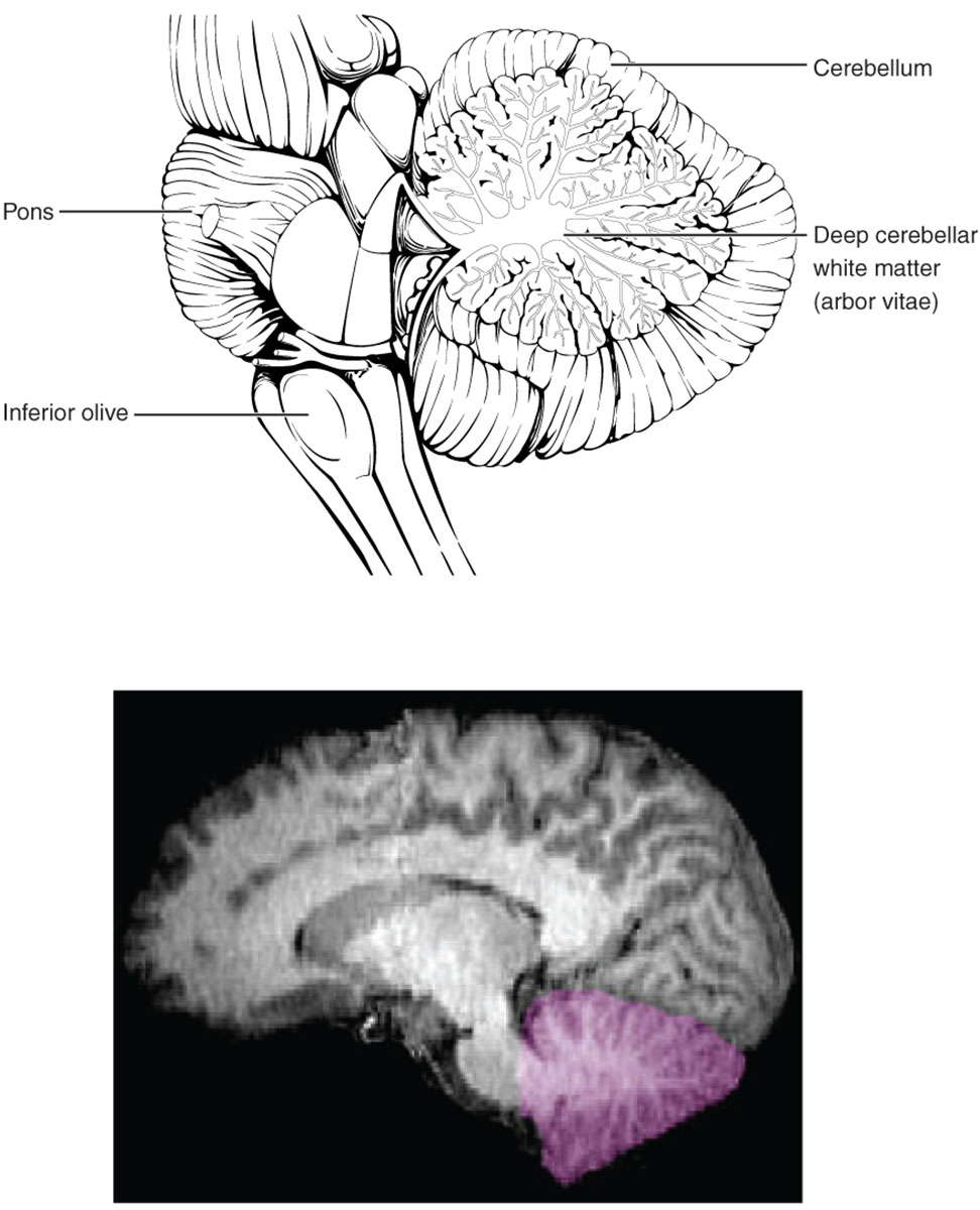
Cerebellum
The cerebellum is located posterior to the brain stem and is responsible for fine-tuning motor activities, maintaining posture, and contributing to cognitive processes like attention and language. Its folded structure, known as folia, increases surface area for neural processing, receiving inputs from the spinal cord and cerebral cortex via the pons and inferior olive.
Pons
The pons serves as a bridge between the midbrain and medulla, relaying signals from the cerebral cortex to the cerebellum through large white matter tracts. It also houses nuclei for cranial nerves involved in facial sensation and movement, supporting respiratory rhythm and arousal.
Inferior olive
The inferior olive, a nucleus in the medulla, provides climbing fiber inputs to the cerebellum, crucial for motor learning and error correction. These fibers synapse on Purkinje cells, influencing cerebellar output to refine movements and adapt to new tasks.
Deep cerebellar white matter (arbor vitae)
The deep cerebellar white matter, resembling a tree-like structure called arbor vitae, consists of myelinated axons that connect cerebellar cortex to deep nuclei and external pathways. This branching pattern facilitates efficient communication between cerebellar regions, enabling coordinated signal transmission for balance and precision.
Anatomical Overview of the Cerebellum
The cerebellum occupies about 10% of brain volume but contains over half of its neurons, underscoring its density and importance. Its position on the brain stem’s posterior surface allows seamless integration with sensory and motor systems.
- The cerebellum is divided into three lobes: anterior, posterior, and flocculonodular, each handling specific aspects of movement and vestibular function.
- Surface folia are separated by fissures, with the primary fissure distinguishing anterior from posterior lobes.
- Deep nuclei—dentate, interposed, and fastigial—serve as output relays, projecting to the thalamus and brain stem.
- Blood supply from the superior cerebellar, anterior inferior cerebellar, and posterior inferior cerebellar arteries ensures metabolic support.
Structural Details of the Pons and Its Connections
The pons forms a prominent bulge in the brain stem, rich in transverse fibers. Its role in connecting higher brain centers to the cerebellum is vital for motor planning.
- Pontocerebellar tracts carry cortical inputs to the cerebellum, facilitating predictive movement adjustments.
- The pontine reticular formation contributes to locomotion and eye movement control via paramedian pontine reticular formation.
- Cranial nerve nuclei within the pons include trigeminal for jaw movement and abducens for lateral gaze.
- Basilar artery branches perfuse the pons, with venous drainage into the petrosal sinuses.
Role and Anatomy of the Inferior Olive
The inferior olive exhibits a folded, olive-shaped appearance, located in the ventral medulla. It provides essential excitatory inputs to the cerebellum for adaptive learning.
- Climbing fibers from the inferior olive wrap around Purkinje cell dendrites, delivering strong synaptic signals for timing and error detection.
- Olivocerebellar tracts cross the midline, ensuring contralateral control of motor corrections.
- GABAergic modulation within the olive regulates firing patterns, synchronized by gap junctions.
- Afferents from the spinal cord and red nucleus inform olivary activity, integrating proprioceptive feedback.
Insights into Deep Cerebellar White Matter
The arbor vitae pattern emerges from myelinated fiber bundles in the cerebellar core. This white matter framework supports rapid neural conduction.
- Axons from Purkinje cells converge on deep nuclei, forming the primary output pathway.
- Superior, middle, and inferior cerebellar peduncles connect the cerebellum to the midbrain, pons, and medulla, respectively.
- Myelin sheaths, produced by oligodendrocytes, enhance signal speed via saltatory conduction.
- Diffusion tensor imaging reveals fiber tract integrity, useful for assessing connectivity.
Imaging Perspectives of the Cerebellum
The accompanying imaging view highlights the cerebellum in purple, demonstrating its appearance in neuroimaging. Such visuals aid in identifying structural details non-invasively.
- MRI techniques like T1-weighted imaging show the arbor vitae as branching hyperintensities due to myelin content.
- Functional MRI detects cerebellar activation during tasks involving coordination and timing.
- The highlighted region emphasizes the vermis, central to axial posture, and hemispheres for appendicular control.
- Volumetric analysis in imaging quantifies cerebellar atrophy in conditions like spinocerebellar ataxias.
Physiological Integration and Functions
The cerebellum processes inputs from multiple sources to refine outputs. Its circuitry ensures smooth, accurate movements through predictive modeling.
- Mossy fibers convey sensory data via granule cells, exciting Purkinje cells in parallel fiber arrays.
- Inhibitory Purkinje output modulates deep nuclei, influencing thalamic and brain stem relays.
- Cerebellar loops with the basal ganglia and cortex support non-motor functions like executive control.
- Neurotransmitters include glutamate for excitation and GABA for inhibition, maintaining circuit balance.
Conclusion
The cerebellum, as depicted in this anatomical illustration and imaging, reveals a highly organized structure essential for motor precision and beyond. With components like the pons, inferior olive, and deep cerebellar white matter working in concert, it exemplifies the brain’s capacity for integration and adaptation. This exploration underscores its significance in neurological function and invites further study into its complex contributions.

