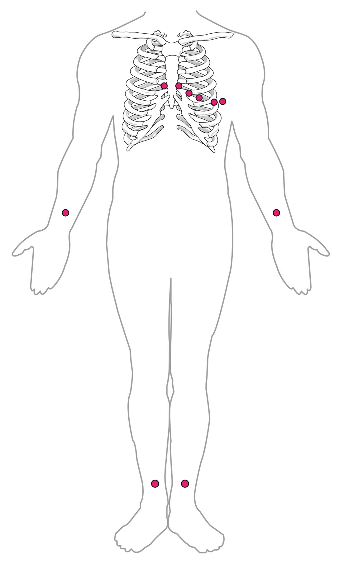
An electrocardiogram (ECG) is a vital tool for assessing heart electrical activity, requiring precise electrode placement for accurate readings. This diagram illustrates the standard placement of the chest electrodes and limb electrodes in a 12-lead ECG, with six electrodes on the chest and four on the limbs, providing a detailed map of cardiac function. Exploring this image enhances understanding of how proper electrode positioning supports effective heart monitoring.
Labelled Parts Explanation
- V1 (4th intercostal space, right sternal border) The V1 electrode, placed in the 4th intercostal space at the right sternal border, records electrical activity from the right ventricle and septum. It helps detect issues like right bundle branch block or septal infarction.
- V2 (4th intercostal space, left sternal border) The V2 electrode, located in the 4th intercostal space at the left sternal border, captures signals from the septum and anterior wall of the left ventricle. It is crucial for identifying anterior myocardial infarction.
- V3 (midway between V2 and V4) The V3 electrode, positioned midway between V2 and V4, records activity from the anterior and septal regions of the left ventricle. It aids in assessing the transition zone between V2 and V4 for ischemic changes.
- V4 (5th intercostal space, midclavicular line) The V4 electrode, placed in the 5th intercostal space at the midclavicular line, monitors the apical region of the left ventricle. It is key for detecting apical infarction or left ventricular hypertrophy.
- V5 (5th intercostal space, anterior axillary line) The V5 electrode, located in the 5th intercostal space at the anterior axillary line, records from the lateral wall of the left ventricle. It helps identify lateral wall ischemia or infarction.
- V6 (5th intercostal space, midaxillary line) The V6 electrode, positioned in the 5th intercostal space at the midaxillary line, captures lateral wall activity of the left ventricle. It is useful for diagnosing lateral myocardial damage.
- RA (right arm) The RA electrode, placed on the right arm, serves as a grounding or reference lead for the limb leads. It contributes to the formation of leads I, II, and III in the standard 12-lead ECG.
- LA (left arm) The LA electrode, located on the left arm, works with the RA and LL electrodes to generate leads I, II, and III. It provides critical data on the heart’s electrical axis and rhythm.
- LL (left leg) The LL electrode, placed on the left leg, acts as a reference point for the limb leads, contributing to leads II, III, and aVF. It helps assess the inferior wall of the heart.
- RL (right leg) The RL electrode, positioned on the right leg, serves as a grounding electrode to reduce electrical interference. It ensures a stable baseline for accurate ECG recordings.
Anatomical Overview of ECG Lead Placement
The standard placement of ECG leads is essential for capturing the heart’s electrical activity accurately. This diagram outlines the strategic positioning that provides a comprehensive view of cardiac function.
- The V1 to V6 chest electrodes map the precordial region, covering the ventricles’ anterior, lateral, and septal areas.
- The RA, LA, LL, and RL limb electrodes form the limb leads, assessing the heart’s frontal plane.
- Proper placement ensures all 12 leads, including precordial and augmented leads, are recorded.
- This configuration allows detection of regional cardiac abnormalities.
Accurate placement is the foundation of reliable ECG interpretation.
Role of Chest Electrodes in ECG Recording
Chest electrodes provide detailed views of the heart’s ventricular activity. Their specific locations enhance diagnostic precision.
- The V1 and V2 electrodes focus on the septum and right ventricle, detecting early conduction issues.
- The V3 and V4 electrodes cover the anterior and apical regions, identifying infarction patterns.
- The V5 and V6 electrodes monitor the lateral wall, revealing lateral ischemia.
- Consistent placement ensures reproducible results across recordings.
These electrodes are vital for localized heart assessment.
Function of Limb Electrodes in ECG Analysis
Limb electrodes contribute to the frontal plane leads, offering a broad view of cardiac electrical activity. Their placement supports axis determination.
- The RA and LA electrodes form lead I, assessing the lateral heart axis.
- The LL electrode, with RA and LA, generates leads II and III, focusing on the inferior wall.
- The RL electrode minimizes noise, stabilizing the ECG baseline.
- This setup provides a three-dimensional perspective of heart function.
Limb leads are essential for rhythm and axis evaluation.
Physiological Importance of Lead Placement
Proper ECG lead placement optimizes the heart’s electrical signal capture. This accuracy supports clinical decision-making.
- The V1 to V6 positions align with cardiac anatomy, maximizing signal strength.
- The RA, LA, LL, and RL leads reflect the heart’s electrical axis in the frontal plane.
- Correct placement reduces artifacts, ensuring clear waveforms.
- This precision aids in diagnosing conditions like arrhythmias or ischemia.
Accurate placement enhances diagnostic reliability.
Clinical Relevance of ECG Lead Positioning
Understanding ECG lead placement is crucial for diagnosing cardiac conditions. These positions are key clinical tools.
- Misplacement of V1 or V2 can mimic right ventricular hypertrophy or infarction.
- The V4 to V6 leads help detect left ventricular strain or lateral wall damage.
- Improper RA or LA placement can alter the electrical axis, leading to misdiagnosis.
- Standardized positioning is critical for serial ECG comparisons.
This knowledge guides effective cardiac monitoring and treatment.
Conclusion
The standard placement of ECG leads diagram provides a detailed guide to the strategic positioning of V1 through V6 chest electrodes and RA, LA, LL, and RL limb electrodes in a 12-lead ECG. By exploring how these placements capture the heart’s electrical activity across the precordial and frontal planes, one gains insight into the importance of accuracy in heart monitoring. This understanding serves as a foundation for studying cardiac diagnostics and addressing related health concerns, encouraging further exploration of ECG techniques and their role in maintaining cardiovascular health.

