The facet joints, also known as zygapophyseal joints, represent crucial articulations between vertebral segments that guide spinal movement while maintaining stability. These specialized synovial joints demonstrate complex biomechanical properties that vary by spinal region, making their understanding essential for medical professionals involved in spine care and surgery.
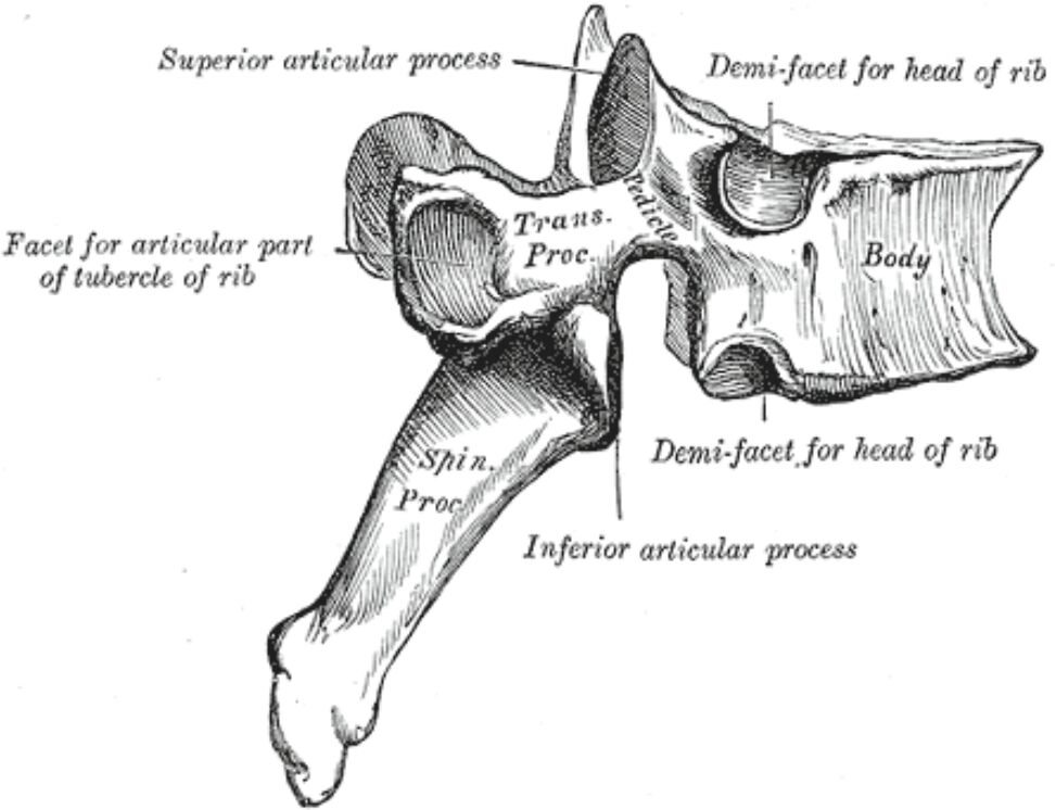
Superior articular process The superior articular process extends upward from the vertebral arch. This process contains a facet joint surface that articulates with the inferior articular process of the vertebra above, creating a sophisticated system for controlled spinal movement.
Transverse Process (Trans. Proc.) The transverse process projects laterally from the vertebral arch. This bony projection serves as an attachment point for muscles and ligaments while also articulating with ribs in the thoracic region.
Spinous Process (Spin. Proc.) The spinous process extends posteriorly from the vertebral arch. It provides attachment points for muscles and ligaments crucial for spinal movement and stability.
Facet for articular part of tubercle of rib This specialized facet surface articulates with the rib tubercle. The joint formed here contributes to the costovertebral joint complex in thoracic vertebrae.
Demi-facet for head of rib The demi-facet represents half of the articular surface for the rib head. When aligned with the corresponding demi-facet of adjacent vertebrae, it forms a complete articular surface for rib attachment.
Inferior articular process The inferior articular process extends downward to articulate with the superior articular process of the vertebra below. Its orientation varies by spinal region to optimize regional biomechanics.
Body The vertebral body represents the primary weight-bearing structure. It provides attachment for intervertebral discs and contains the demi-facets for rib articulation in thoracic vertebrae.
Facet Joint Architecture and Function
The facet joints exemplify nature’s engineering in spinal design. Their complex architecture enables controlled movement while providing stability through multiple planes. These joints demonstrate remarkable adaptation to regional biomechanical demands throughout the spine.
Biomechanical Considerations
The orientation of facet joints varies by spinal region to optimize function. This variation allows for different movement patterns while maintaining structural integrity and protecting neural elements.
Clinical Significance
Diagnostic Approaches
Modern imaging techniques reveal important details about facet joints:
- CT imaging for bone architecture
- MRI for soft tissue evaluation
- Dynamic studies for movement patterns
- 3D reconstruction for surgical planning
Pathological Conditions
Common facet joint conditions include:
- Facet arthropathy
- Synovial cysts
- Degenerative changes
- Traumatic injuries
Advanced Anatomical Relationships
Neurovascular Considerations
Critical structures near facet joints include:
- Spinal nerve roots
- Medial branches
- Vascular supply
- Ligamentous attachments
Muscular Interactions
Multiple muscle groups influence joint function:
- Deep spinal muscles
- Multifidus system
- Rotator muscles
- Intersegmental muscles
Modern Treatment Applications
Conservative Management
Non-surgical approaches include:
- Facet injections
- Medial branch blocks
- Physical therapy
- Exercise modification
Surgical Interventions
When necessary, surgical options include:
- Facet fusion
- Decompression procedures
- Total joint replacement
- Minimally invasive techniques
Future Developments
Emerging Technologies
Current research explores:
- Novel implant designs
- Biological treatments
- Regenerative approaches
- Advanced imaging methods
- Facet Joint Anatomy: A Complete Guide for Medical Professionals
- Understanding Spinal Facet Joints: Structure and Function
- Comprehensive Analysis of Vertebral Articular Processes
- Facet Joint Architecture: Clinical Perspectives
- Essential Guide to Spinal Facet Joint Anatomy

