Deep Vein Thrombosis (DVT) is a serious vascular condition characterized by the formation of a blood clot (thrombus) within a deep vein, predominantly occurring in the lower extremities such as the calf or thigh. The clinical presentation of this condition is often visually distinct, manifesting as significant asymmetry between the limbs due to fluid retention and inflammation. The image provided illustrates a classic presentation of a right leg DVT, highlighting the contrast in size and skin tone compared to the unaffected left leg, serving as a critical example of why early visual recognition is vital for preventing severe complications.
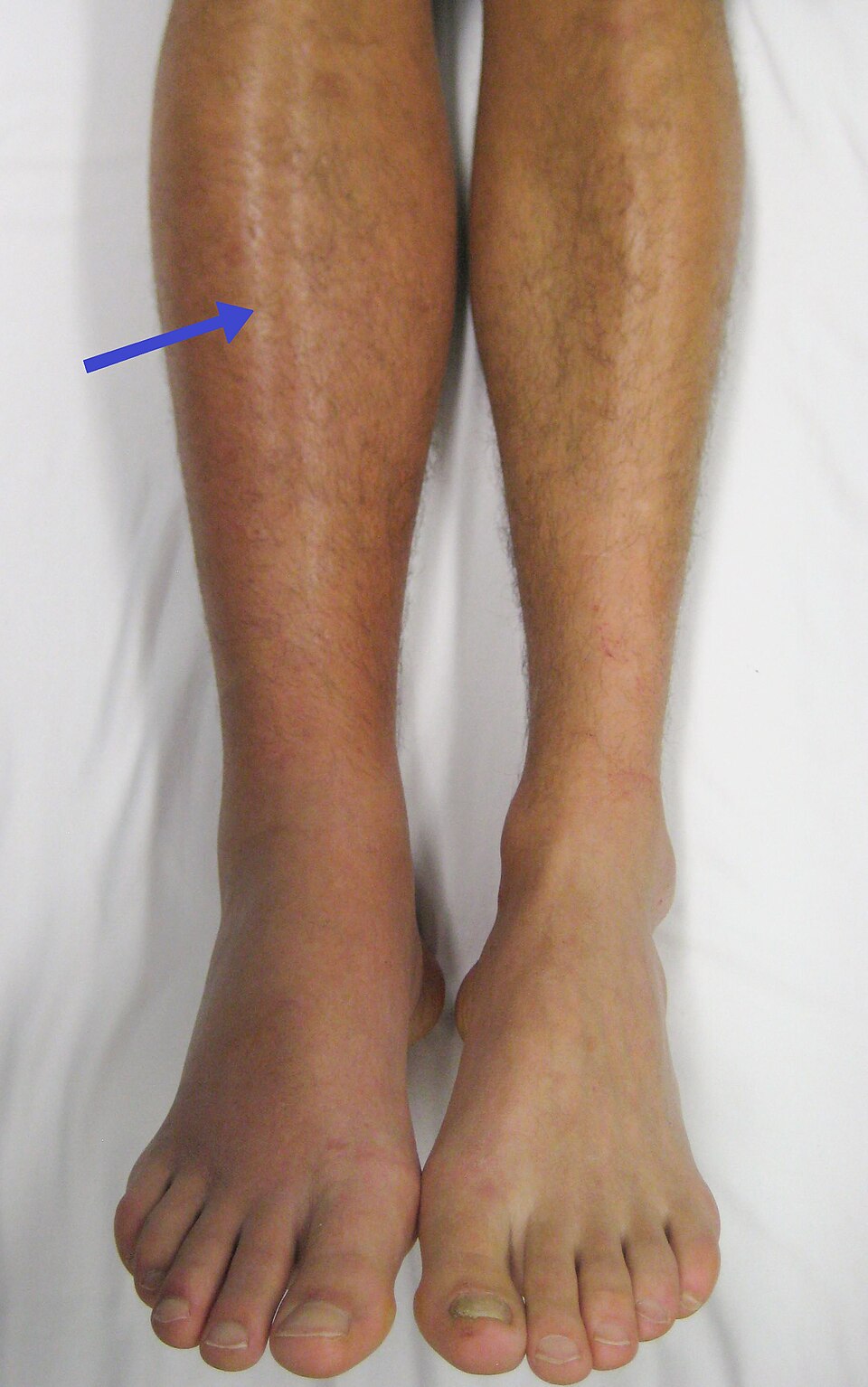
Blue Arrow: The arrow points directly to the right calf, highlighting a distinct area of unilateral edema (swelling) and erythema (redness). This visible enlargement compared to the opposite leg suggests that a blockage in the deep venous system is preventing proper blood drainage, leading to fluid accumulation and tissue inflammation.
Understanding the Clinical Presentation of DVT
Deep Vein Thrombosis is a condition that demands immediate medical attention due to its potential to evolve into life-threatening emergencies. The thrombus usually forms in the deep veins of the leg—such as the popliteal, femoral, or iliac veins—often due to a combination of factors known as Virchow’s Triad: stasis of blood flow, endothelial injury (damage to the vein wall), and hypercoagulability (increased tendency of the blood to clot). When a clot forms, it acts as a physical barrier, obstructing the return of deoxygenated blood to the heart.
This obstruction leads to an increase in hydrostatic pressure within the vein, forcing fluid out of the vascular space and into the surrounding interstitial tissues. This process results in the swelling seen in the image. Furthermore, the inflammatory response to the clot causes the release of various mediators, leading to the redness (erythema) and warmth that are frequently palpable upon examination. While some cases of DVT are “silent” and present no symptoms, symptomatic cases usually present with a specific set of warning signs.
Common signs and symptoms associated with lower extremity DVT include:
- Swelling in one leg (rarely both legs simultaneously).
- Pain or tenderness in the leg, often starting in the calf and feeling like cramping or soreness.
- Red or discolored skin on the leg.
- A feeling of warmth in the affected leg.
- Visible surface veins (collateral veins) becoming more prominent.
It is crucial to differentiate DVT from other causes of leg pain and swelling, such as cellulitis, muscle strains, or lymphedema. However, the stakes with DVT are significantly higher. If a portion of the clot breaks loose, it becomes an embolus and can travel through the bloodstream to the lungs, causing a Pulmonary Embolism (PE). A PE can block blood flow to the lungs, lowering oxygen levels and potentially causing heart failure, making DVT a diagnosis that cannot be missed.
Pathophysiology and Risk Factors
The physiological mechanism behind DVT centers on the disruption of normal hemostasis. Under normal conditions, the body maintains a delicate balance between clot formation (to stop bleeding) and clot dissolution (fibrinolysis). In DVT, this balance tips toward coagulation. Risk factors are numerous and include prolonged immobility (such as long flights or bed rest), recent surgery (especially orthopedic procedures like hip or knee replacements), trauma, active cancer, and hormonal therapies like oral contraceptives.
Anatomically, the deep veins are surrounded by muscle. The contraction of the calf muscles, often called the “second heart,” pumps blood against gravity back toward the heart. When a patient is immobile, this pump is inactive, leading to venous stasis—a primary driver for clot formation. Once the thrombus is established, it can propagate, extending further up the leg. The body’s natural response involves inflammation, which explains the redness and heat noted in the patient’s right leg in the image.
Diagnosis and Therapeutic Approaches
Diagnosing Deep Vein Thrombosis typically begins with a physical examination and a pre-test probability assessment, such as the Wells Score. If DVT is suspected, the gold standard for diagnosis is a duplex ultrasonography. This non-invasive imaging test uses sound waves to visualize the blood flow in the veins and can detect the presence of a clot. In some cases, a D-dimer blood test is used; elevated levels of D-dimer, a protein fragment produced when a blood clot dissolves, suggest the presence of a thrombotic event, though it is not specific to DVT alone.
Treatment focuses on preventing the clot from getting bigger, preventing it from breaking loose, and reducing the chances of another clot forming. Anticoagulant medications, often referred to as “blood thinners,” are the cornerstone of therapy. These include injectables like low molecular weight heparin or oral medications such as warfarin and direct oral anticoagulants (DOACs). In severe cases, thrombolytics (clot busters) might be used to dissolve the clot directly, or a filter may be inserted into the inferior vena cava to catch clots before they reach the lungs.
Conclusion
Recognizing the physical manifestations of a Deep Vein Thrombosis, such as the pronounced swelling and redness depicted in the right leg of the patient, is essential for timely medical intervention. These visual cues serve as early warnings of a potentially fatal condition. By understanding the underlying anatomy and physiological risks, patients and healthcare providers can act quickly to diagnose the issue and initiate anticoagulant therapy, thereby significantly reducing the risk of complications and ensuring better long-term health outcomes.

