The provided image offers a clear and concise comparison between a normally positioned placenta and a condition known as placenta previa. This condition arises when an embryo implants too close to the cervical opening, leading to the placenta partially or completely covering the cervix. This distinction is critical in obstetrics, as placenta previa can significantly impact the course of pregnancy and delivery.
Anatomical and Pathological Labels Explained
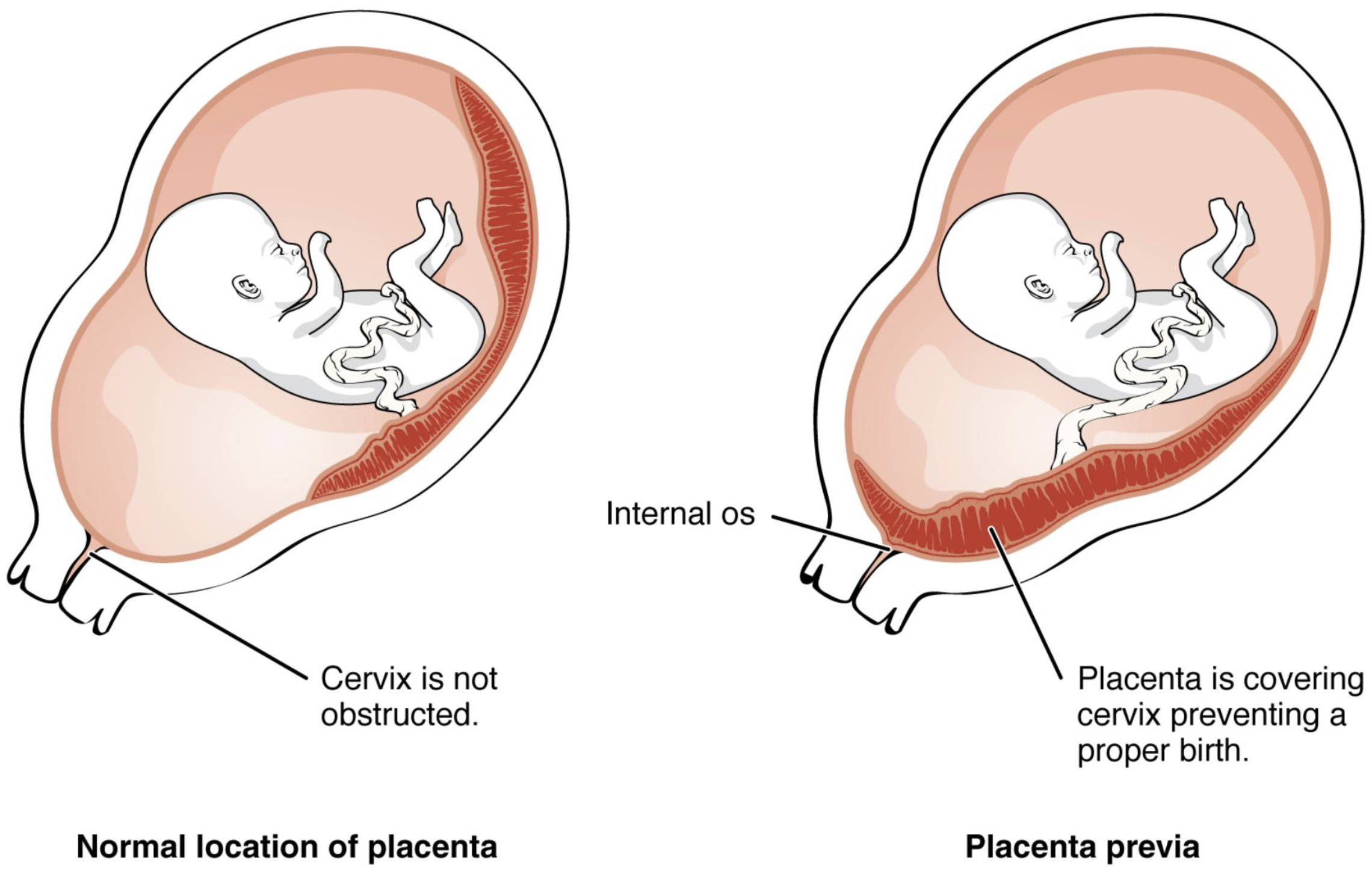
The diagram illustrates two distinct scenarios, one representing a healthy placental position and the other depicting placenta previa. Understanding these labels is fundamental to grasping the implications of this condition.
Cervix is not obstructed: This label indicates a healthy pregnancy where the cervix, the opening to the uterus, remains clear. In this normal scenario, the placenta is positioned higher in the uterus, allowing for an unobstructed vaginal birth.
Normal location of placenta: This caption refers to the typical and safe position of the placenta within the uterus, usually in the upper segment. A normally located placenta ensures that the cervical canal is clear, facilitating a natural delivery process.
Internal os: This refers to the internal opening of the cervix, which connects the cervical canal to the uterine cavity. In cases of placenta previa, the placenta covers this crucial opening, impeding the baby’s passage during birth.
Placenta is covering cervix preventing a proper birth: This label highlights the central issue in placenta previa, where the placenta obstructs the cervical opening. This obstruction poses a significant risk during labor, as it can lead to severe bleeding and necessitates a caesarean section for safe delivery.
Placenta previa: This term denotes a condition where the placenta implants in the lower uterine segment, either partially or completely covering the internal os of the cervix. This abnormal implantation can lead to various complications during pregnancy and childbirth.
Delving Deeper into Placenta Previa
Placenta previa is a significant obstetric complication that arises from the abnormal implantation of the placenta in the lower uterine segment, consequently covering the internal os of the cervix. This condition can be categorized based on the degree to which the placenta covers the cervical opening:
- Complete previa: The placenta entirely covers the internal os.
- Partial previa: The placenta partially covers the internal os.
- Marginal previa: The placenta extends to the edge of the internal os but does not cover it.
- Low-lying placenta: The placenta is implanted in the lower uterine segment but its edge is within 2 cm of the internal os without covering it.
The primary concern with placenta previa is the risk of antepartum hemorrhage, which is vaginal bleeding after 20 weeks of gestation. As the lower uterine segment thins and dilates during the third trimester or labor, the placenta, being less elastic, can separate from the uterine wall, leading to bleeding. This bleeding is typically painless, bright red, and can be severe, potentially endangering both the mother and the fetus.
The diagnosis of placenta previa is usually made during routine prenatal ultrasound examinations. If identified early in pregnancy, many cases of low-lying placenta or marginal previa may resolve spontaneously as the uterus grows and the placental position appears to “migrate” away from the cervix, a phenomenon known as placental trophism or migration. However, if the placenta remains covering the cervix as term approaches, a caesarean section becomes the safest mode of delivery to prevent hemorrhage and ensure the well-being of the mother and baby. Management typically involves close monitoring, activity restrictions, and, in some cases, hospitalization to manage potential bleeding episodes.
The implications of placenta previa extend beyond just the mode of delivery. It can also be associated with other pregnancy complications, such as preterm birth, fetal growth restriction, and an increased risk of postpartum hemorrhage due to the lower uterine segment’s reduced ability to contract effectively after delivery. Understanding the anatomical differences, as clearly depicted in the diagram, is crucial for timely diagnosis and appropriate management to ensure optimal outcomes for both mother and child in cases of placenta previa.

