The pituitary gland, often referred to as the “master gland,” is a small but vital structure located in the sella turcica of the sphenoid bone, just below the brain. This diagram illustrates the normal anatomy of the pituitary gland and the changes caused by a pituitary tumor, highlighting its potential to affect vision due to its proximity to the optic chiasm. Understanding these illustrations provides key insights into the gland’s function and the clinical implications of tumor growth.
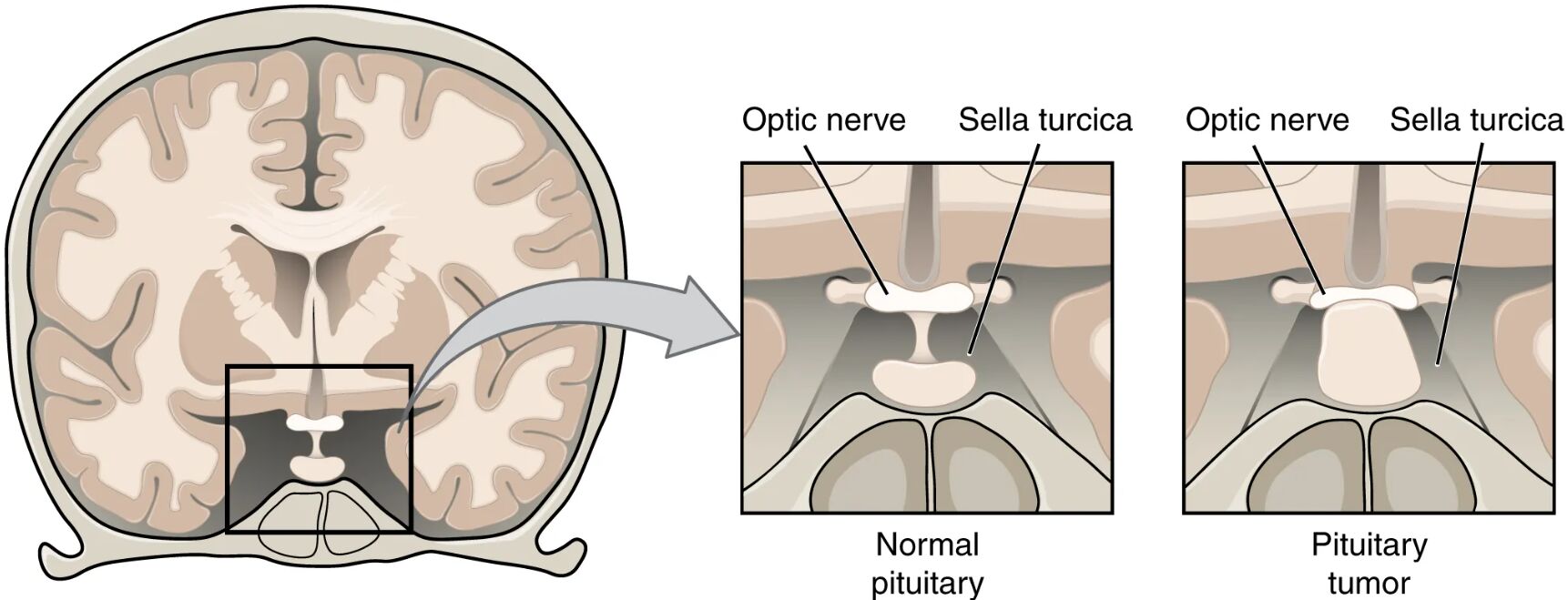
Optic nerve: The optic nerve carries visual information from the retina to the brain, crossing at the optic chiasm where fibers from the nasal halves of each retina converge. A pituitary tumor pressing on this area can disrupt these fibers, leading to visual field defects.
Sella turcica: The sella turcica is a saddle-shaped depression in the sphenoid bone that houses the pituitary gland, providing a protective bony enclosure. Expansion of a tumor within this space can compress surrounding structures, including the optic chiasm.
Normal pituitary: The normal pituitary gland is a small, pea-sized structure that secretes hormones like growth hormone, thyroid-stimulating hormone, and adrenocorticotropic hormone to regulate various bodily functions. It fits snugly within the sella turcica without exerting pressure on adjacent nerves.
Pituitary tumor: A pituitary tumor is an abnormal growth that can enlarge the gland, potentially compressing the optic chiasm and causing symptoms like tunnel vision. These tumors may be benign or functional, producing excess hormones that affect metabolism or growth.
Anatomical Overview of the Pituitary Gland
The pituitary gland is centrally located at the base of the brain, playing a pivotal role in hormonal regulation. This diagram offers a clear view of its position and the potential impact of a tumor.
- The optic nerve’s path near the pituitary makes it vulnerable to compression by a growing tumor.
- The sella turcica serves as a natural cradle, stabilizing the gland while allowing hormone secretion into the bloodstream.
- The normal pituitary maintains a balanced hormone output, influencing organs like the thyroid and adrenal glands.
- A pituitary tumor disrupts this balance, potentially leading to hormonal imbalances or neurological symptoms.
Its strategic location underscores its importance in overall endocrine health.
Effects of Pituitary Tumors
The diagram illustrates how a pituitary tumor alters the gland’s structure and impacts nearby anatomy. This visual aid helps in understanding the associated symptoms.
- A pituitary tumor can expand beyond the sella turcica, pressing on the optic nerve and causing visual disturbances.
- Tunnel vision results from damage to the optic chiasm, where the tumor interrupts peripheral vision fibers.
- The normal pituitary’s compact size contrasts with the tumor’s potential to distort surrounding tissues.
- Hormonal overproduction from functional tumors may lead to conditions like acromegaly or Cushing’s disease.
Early detection through MRI or CT scans is essential for managing these effects.
Clinical Symptoms and Diagnosis
Pituitary tumors present a range of symptoms due to their location and hormonal activity. Recognizing these signs aids in timely diagnosis.
- Compression of the optic nerve by a pituitary tumor often manifests as vision loss, starting with peripheral fields.
- Headaches may arise from the tumor’s pressure within the confined sella turcica.
- Excess hormone secretion from the normal pituitary can be disrupted, leading to fatigue or weight changes.
- Blood tests and imaging help differentiate between benign and malignant growths, guiding treatment plans.
Endocrinologists use hormone level assessments to confirm tumor activity.
Treatment and Management
Managing a pituitary tumor involves addressing both the tumor and its effects on the optic nerve and hormonal balance. Treatment options vary based on tumor size and symptoms.
- Surgical removal via transsphenoidal surgery is common for large tumors compressing the optic nerve.
- Radiation therapy may be used for residual tumor tissue within the sella turcica.
- Medications can control hormone overproduction if the normal pituitary function is altered.
- Regular monitoring ensures the tumor does not recur, preserving vision and endocrine health.
A multidisciplinary approach, including neurosurgeons and endocrinologists, optimizes outcomes.
Conclusion
This diagram of the pituitary gland and a pituitary tumor provides a clear illustration of its anatomy and the potential complications arising from tumor growth. The optic nerve, sella turcica, normal pituitary, and tumor dynamics highlight the gland’s critical role and the need for prompt intervention. This understanding equips individuals with the knowledge to recognize symptoms and seek appropriate care for effective management.

