The parathyroid glands, small yet vital endocrine organs, are nestled on the posterior surface of the thyroid gland, playing a key role in calcium homeostasis. This article delves into their anatomical structure as seen from the posterior view, offering a clear perspective on their location and relationship with surrounding neck structures.
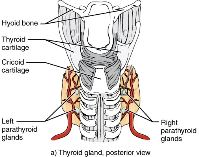
Hyoid bone The hyoid bone is positioned above the thyroid gland, providing structural support to the neck. It anchors the tongue and larynx, indirectly stabilizing the parathyroid glands’ upper region.
Thyroid cartilage The thyroid cartilage, known as the Adam’s apple, protects the larynx and supports vocal cord function. It forms a protective boundary around the upper thyroid where the parathyroid glands reside.
Cricoid cartilage The cricoid cartilage forms the base of the larynx, located below the thyroid cartilage. It offers additional stability to the lower thyroid and parathyroid gland area.
Left parathyroid glands The left parathyroid glands are embedded on the left posterior surface of the thyroid, secreting parathyroid hormone (PTH) to regulate blood calcium. Their small size and strategic location ensure effective calcium control.
Right parathyroid glands The right parathyroid glands mirror the left, situated on the right posterior thyroid surface, releasing PTH to maintain calcium levels. Their bilateral placement supports balanced endocrine activity.
Anatomical Overview of the Parathyroid Glands
The posterior view highlights the parathyroid glands’ integration with the thyroid. This perspective reveals their anatomical context within the neck.
- The hyoid bone provides upper support, anchoring the thyroid and parathyroid region.
- The thyroid cartilage shields the larynx, offering protection to the upper glands.
- The cricoid cartilage stabilizes the lower thyroid, aiding parathyroid positioning.
- The left parathyroid glands regulate calcium on the left side of the thyroid.
- The right parathyroid glands ensure symmetrical calcium homeostasis on the right.
Role of Surrounding Structures
The cartilages and thyroid gland create a supportive environment for the parathyroid glands. Their anatomical relationships enhance gland function.
- The hyoid bone’s position supports neck mobility, indirectly benefiting the glands.
- The thyroid cartilage’s protective role safeguards the parathyroid glands from injury.
- The cricoid cartilage provides a stable base for the lower thyroid region.
- The thyroid gland’s posterior surface serves as a natural embedding site for the glands.
- This structural support ensures efficient PTH secretion and distribution.
Physiological Functions of the Parathyroid Glands
The parathyroid glands regulate blood calcium levels through PTH secretion. This function is crucial for bone health and neuromuscular activity.
- PTH increases calcium by stimulating bone resorption and kidney reabsorption.
- The glands respond to low calcium levels, releasing PTH to restore balance.
- The left and right glands work in unison to maintain systemic calcium homeostasis.
- Their posterior location on the thyroid allows close proximity to blood supply.
- This regulation prevents conditions like hypocalcemia or hypercalcemia.
Clinical Relevance and Imaging Techniques
Understanding the posterior anatomy of the parathyroid glands aids in clinical practice. This knowledge is essential for diagnosis and treatment.
- The glands’ location on the thyroid requires precise imaging for identification.
- The hyoid and cricoid cartilages guide surgical approaches to avoid damage.
- The thyroid cartilage’s prominence helps in palpating the neck for abnormalities.
- Ultrasound or MRI can assess the glands’ size and position.
- Conditions like hyperparathyroidism may necessitate gland evaluation.
The parathyroid glands’ posterior placement on the thyroid gland, supported by the hyoid, thyroid, and cricoid cartilages, underscores their critical role in calcium regulation. This anatomical arrangement ensures effective hormone production and distribution, offering valuable insights for clinical management and understanding endocrine health.

