Observing Paramecium under the microscope reveals the dynamic life of one of biology’s most iconic single-celled eukaryotes, showcasing complex behaviors and specialized organelles within a single cell. This slipper-shaped ciliate, commonly found in freshwater environments, serves as an exceptional model organism for studying ciliary motility, nuclear dimorphism, osmoregulation, and intracellular digestion. The microscopic view highlights key structures that allow Paramecium to swim, feed, and maintain homeostasis, providing students, educators, and researchers with a clear window into eukaryotic cell biology that has direct parallels to processes occurring in human cells, particularly in ciliary function and sensory signaling.
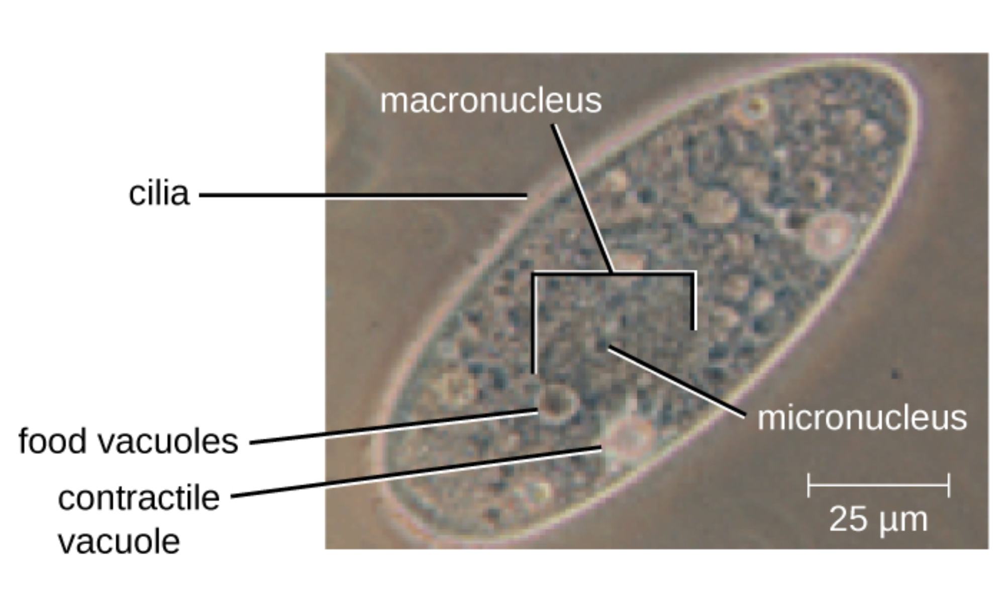
Macronucleus is the large, irregular, often kidney-shaped structure visible in the central region of the cell. It is highly polyploid and governs all vegetative functions, including metabolism, protein synthesis, and daily cellular activities through its abundant DNA copies.
Micronucleus is the smaller, more compact nucleus typically located near the macronucleus. It functions primarily as the germline nucleus, preserving the complete diploid genome for sexual reproduction via conjugation and maintaining genetic stability across generations.
Cilia are the numerous fine, hair-like projections covering the cell surface, visible as a fuzzy outline in the microscopic image. These motile organelles beat in coordinated waves to propel the organism through water and generate feeding currents that direct particles toward the oral region.
Food vacuoles are the circular, membrane-bound structures scattered throughout the cytoplasm. They contain ingested food particles at various stages of digestion and circulate within the cell as enzymes break down nutrients before waste is expelled.
Contractile vacuole is the clear, spherical organelle often visible near the cell periphery. It collects excess water from the cytoplasm via radiating canals and rhythmically contracts to expel fluid, maintaining osmotic balance in the hypotonic freshwater habitat of Paramecium.
25 μm scale bar provides essential size reference, confirming that Paramecium is a relatively large protist, easily observable under standard light microscopy and suitable for detailed live observation in educational and research settings.
Live Microscopic Observation of Paramecium
When viewed under a light microscope, Paramecium appears as an active, elongated cell constantly changing direction through rapid ciliary beating. The macronucleus is often the most prominent internal feature, while food vacuoles can be seen circulating slowly. Contractile vacuoles fill and collapse at regular intervals, demonstrating active osmoregulation. The constant motion of cilia creates visible currents around the cell, highlighting the organism’s sophisticated sensory and motor capabilities even at the unicellular level.
Key Structural Features Revealed Under the Microscope
The microscopic image clearly shows the characteristic slipper shape of Paramecium with its anterior end slightly more pointed than the posterior. Cilia cover the entire surface, giving the cell a fuzzy appearance. The large macronucleus stands out due to its size and staining properties, while the smaller micronucleus is often visible nearby. Food vacuoles appear as distinct spherical inclusions, and the contractile vacuole is identifiable by its clear appearance and periodic pulsation, features that make Paramecium an ideal subject for live-cell observation.
Ciliary Motility and Behavior
Cilia enable Paramecium to swim at speeds up to 1 mm per second while allowing precise control of direction. When encountering obstacles or unfavorable chemicals, the cell can reverse its ciliary beat, causing it to back up and change course. This avoidance behavior is regulated by calcium influx and membrane potential changes, providing a simple yet powerful model for understanding sensory transduction and motor control in eukaryotes.
- Ciliary beating creates both propulsion and feeding currents.
- Metachronal waves ensure smooth, coordinated movement.
- Calcium signaling controls ciliary reversal for avoidance responses.
These mechanisms have direct relevance to human ciliopathies affecting lungs, kidneys, and brain function.
Nuclear Dimorphism in Action
Paramecium’s dual nuclear system is one of its most fascinating features. The large macronucleus handles daily operations, while the micronucleus remains transcriptionally silent during normal growth but plays a crucial role during sexual conjugation. Under the microscope, the size difference between the two nuclei is readily apparent, illustrating how the organism separates somatic and germline functions within a single cell.
Osmoregulation and Contractile Vacuoles
Living in freshwater, Paramecium constantly gains water by osmosis. The contractile vacuole system counters this by collecting and expelling excess fluid. In live observations, the vacuole can be seen filling gradually before suddenly collapsing, demonstrating an elegant solution to cellular volume regulation that parallels ion transport mechanisms in more complex organisms.
Feeding and Digestion Processes
Paramecium feeds primarily on bacteria and small organic particles. Cilia in the oral groove create currents that sweep food into the cytostome, where it is packaged into food vacuoles. These vacuoles mature as they move through the cell, becoming acidic and receiving digestive enzymes. The presence of multiple food vacuoles at different stages is often visible under the microscope, providing a clear view of intracellular digestion in action.
Paramecium as an Educational and Research Tool
Paramecium is widely used in biology classrooms because its large size and active behavior make it easy to observe under standard microscopes. Students can watch live swimming, feeding, and contractile vacuole activity in real time. In research laboratories, it continues to serve as a model for studying ciliary biology, genome organization, and cellular responses to environmental stimuli, with findings frequently informing our understanding of human diseases.
Conclusion: The Enduring Educational Value of Paramecium
The microscopic view of Paramecium beautifully captures the complexity and elegance of a single-celled organism performing sophisticated tasks. From the prominent macronucleus and active cilia to the pulsing contractile vacuole and circulating food vacuoles, every visible feature illustrates fundamental principles of eukaryotic cell biology. As both an educational tool and a research model, Paramecium continues to inspire new generations of students and scientists, bridging basic observation with advanced insights into cellular function and human health.

