Paramecium is a classic single-celled ciliate that has served as a foundational model organism in cell biology, genetics, and physiology for more than a century. Its complex internal organization, visible in detailed diagrams like the one presented, demonstrates how a unicellular eukaryote can perform sophisticated functions including coordinated motility, feeding, digestion, osmoregulation, and even primitive sexual reproduction. Belonging to the Chromalveolata supergroup, Paramecium continues to provide valuable insights into conserved eukaryotic processes, with particular relevance to human ciliopathies, cellular signaling, and the evolution of multicellularity.
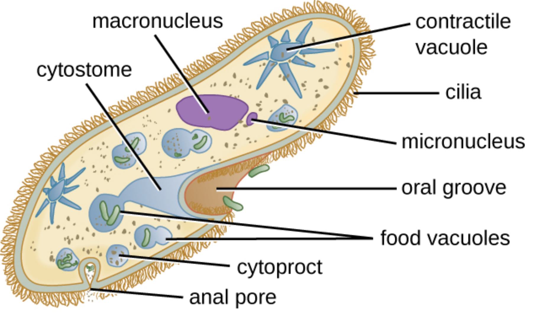
Macronucleus is the large, bean-shaped nucleus prominently displayed in the central region of the cell. It is highly polyploid and controls all vegetative functions, including metabolism and protein synthesis, by maintaining multiple copies of the active genome.
Micronucleus is the small, compact nucleus located near the macronucleus. It functions as the germline nucleus, preserving the complete diploid genome for sexual reproduction through conjugation and ensuring genetic stability across generations.
Contractile vacuole is the star-shaped organelle with radiating canals located toward the anterior and posterior ends. It actively collects excess water from the cytoplasm and expels it through a pore to maintain osmotic balance in the hypotonic freshwater environment inhabited by Paramecium.
Cilia are the numerous fine projections covering the entire cell surface. They beat in coordinated metachronal waves to propel the organism through water and generate currents that direct food particles toward the oral region.
Cytostome is the cell mouth opening located at the base of the oral groove. It serves as the entry point where food particles are engulfed into forming food vacuoles for intracellular digestion.
Oral groove is the shallow, funnel-shaped depression running along one side of the cell. It channels water currents created by ciliary beating, concentrating bacteria and organic debris for efficient feeding.
Food vacuoles are spherical membrane-bound compartments containing ingested food at different stages of digestion. They circulate through the cytoplasm, undergoing acidification and enzymatic breakdown before waste is expelled.
Cytoproct is the posterior opening also known as the cell anus. It allows the release of indigestible residues from mature food vacuoles after digestion is completed.
Anal pore refers to the same structure as the cytoproct, the site where undigested material is egested from the cell into the surrounding environment.
Overall Cellular Architecture of Paramecium
Paramecium exhibits a highly organized body plan despite being unicellular. The cell is enclosed by a pellicle, a flexible yet supportive layer composed of the plasma membrane, alveolar sacs, and underlying epiplasm. Beneath this lies a layer of ectoplasm rich in basal bodies from which cilia emerge in precise longitudinal rows. The endoplasm contains the majority of organelles, including the nuclei, vacuoles, and mitochondria. This polarized structure, with distinct anterior and posterior ends, supports directed locomotion and feeding.
Nuclear Dimorphism and Genetic Regulation
Paramecium displays nuclear dimorphism, possessing both a macronucleus for daily functions and a micronucleus for reproduction. The macronucleus is transcriptionally active with amplified gene copies, while the micronucleus remains transcriptionally silent during vegetative growth but undergoes meiosis during conjugation. This separation allows the organism to maintain a stable germline while supporting high levels of somatic gene expression, a feature that has made Paramecium a key model for studying genome organization and epigenetic regulation.
Ciliary Motility and Sensory Behavior
The dense covering of cilia enables Paramecium to swim rapidly and respond to environmental stimuli. Ciliary beating is coordinated through calcium-dependent mechanisms, allowing the organism to reverse direction when encountering unfavorable conditions. This avoidance behavior, along with chemotaxis and mechanosensation, demonstrates sophisticated sensory integration in a single cell and provides insights into human ciliary disorders known as ciliopathies.
- Cilia generate both propulsive force for swimming and feeding currents.
- Basal body organization ensures precise alignment and coordination.
- Membrane potential changes regulate ciliary reversal.
Studies on Paramecium cilia have advanced understanding of diseases affecting motile cilia in the respiratory tract and brain.
Feeding and Digestive System
Paramecium is a heterotrophic predator that primarily consumes bacteria. The oral groove funnels particles to the cytostome, where they are packaged into food vacuoles. These vacuoles mature as they move through the cell, fusing with lysosomes for digestion. The cytoproct then expels waste, completing an efficient intracellular digestive cycle that parallels aspects of phagocytosis in immune cells.
Osmoregulation by Contractile Vacuoles
In freshwater environments, Paramecium faces constant water influx by osmosis. The contractile vacuoles, with their complex canal systems, collect and expel excess water in rhythmic contractions. This energy-dependent process maintains cellular volume and ion balance, serving as an excellent model for studying membrane transport and osmoregulatory mechanisms conserved in more complex organisms.
Paramecium as a Biomedical Research Model
Beyond its educational value, Paramecium has contributed to major discoveries in genetics, including cytoplasmic inheritance and genome rearrangement. Its transparency, large size, and ease of cultivation make it ideal for live-cell imaging and genetic manipulation. Current research uses Paramecium to study ciliary biogenesis, ion channel function, and cellular responses to environmental stress, with findings often translating to human health applications.
Educational and Practical Significance
Paramecium remains a staple in biology classrooms worldwide due to its visible organelles and dynamic behaviors under the microscope. Its structure illustrates key eukaryotic features such as membrane-bound organelles, cytoskeletal organization, and specialized feeding structures. In research laboratories, it supports high-throughput screening and serves as a safe, non-pathogenic model for exploring fundamental cellular processes.
Conclusion: The Enduring Value of Paramecium Studies
The detailed anatomy of Paramecium, clearly illustrated in the diagram, reveals a remarkably sophisticated unicellular organism capable of complex behaviors and precise physiological control. From its dual nuclei and contractile vacuoles to the array of cilia powering motility and feeding, every feature demonstrates optimized solutions to the challenges of single-celled life. As a model organism, Paramecium continues to illuminate conserved biological principles with direct relevance to human medicine, particularly in the fields of ciliary biology and cellular physiology, ensuring its place as a cornerstone of biological education and research.

