The optic chiasm is a critical junction in the visual system where nerve fibers from the retina partially cross, organizing visual information from both eyes for brain processing. This inferior view diagram illustrates how contralateral and ipsilateral visual field data are segregated, highlighting the pathway that ensures a unified visual perception.
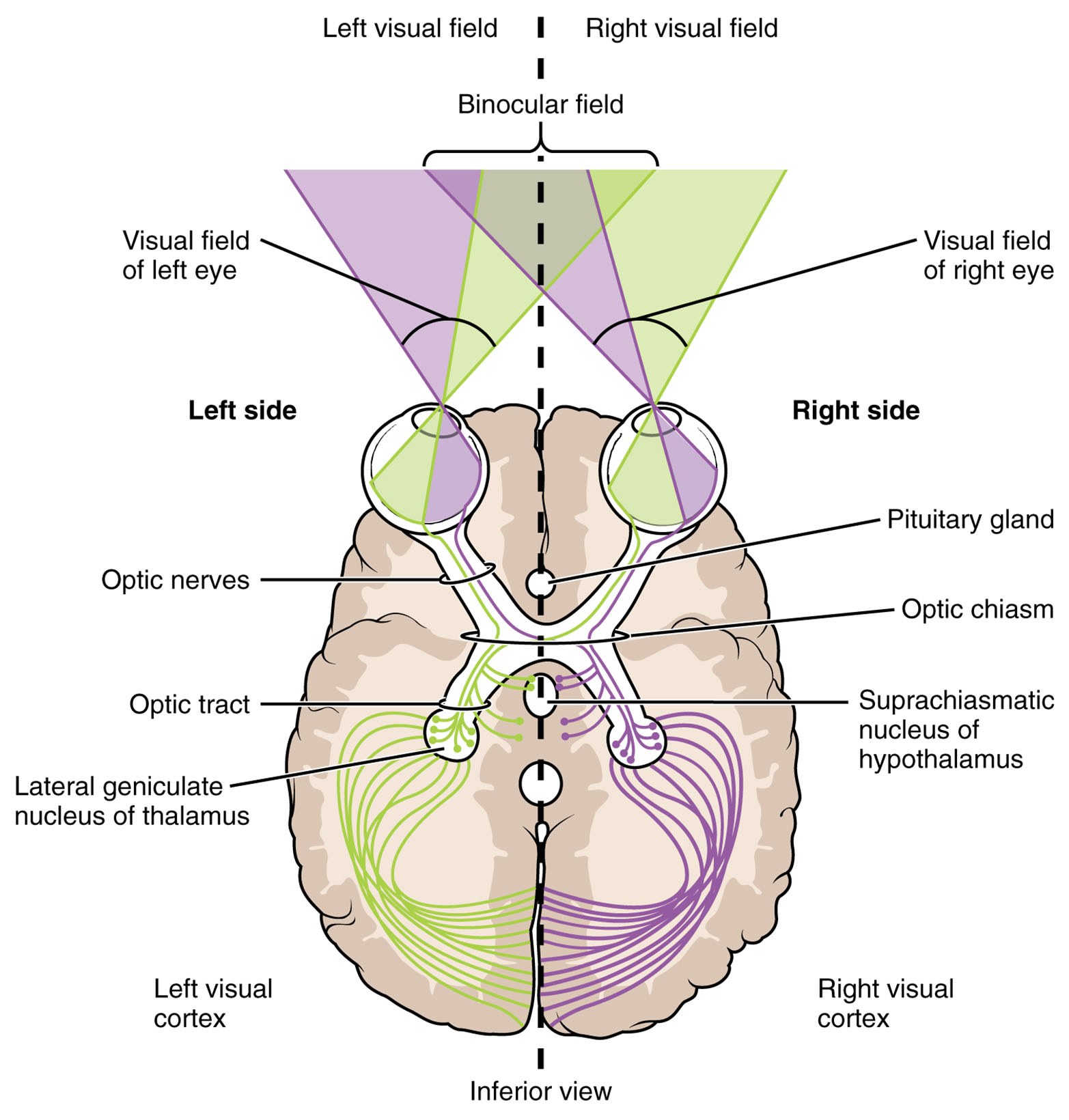
Optic nerve (II) The optic nerve (II) carries visual information from the retina of each eye to the optic chiasm. It contains axons of ganglion cells, transmitting light-induced signals for further processing in the brain.
Optic chiasm The optic chiasm is the point where some optic nerve fibers cross to the opposite side, segregating visual field information. It ensures that the left and right visual fields are represented on both sides of the brain.
Optic tract The optic tract carries nerve fibers from the optic chiasm to the lateral geniculate nucleus, containing a mix of crossed and uncrossed retinal inputs. It relays visual data for processing in higher visual centers.
Lateral geniculate nucleus (LGN) The lateral geniculate nucleus (LGN), part of the thalamus, receives input from the optic tract and processes visual information. It organizes the signals before sending them to the visual cortex.
Visual cortex The visual cortex, located in the occipital lobe, interprets the processed visual signals from the LGN. It constructs the conscious perception of the visual field based on the segregated inputs.
Nasal retina The nasal retina, the inner half of each retina, detects the temporal (lateral) visual field and its fibers cross at the optic chiasm. This crossing allows the brain to integrate visual information from both eyes’ outer fields.
Temporal retina The temporal retina, the outer half of each retina, perceives the nasal (medial) visual field and its fibers remain ipsilateral. These uncrossed fibers contribute to the same side’s brain representation.
Anatomy of the Optic Chiasm
The optic chiasm serves as a pivotal structure where visual pathways reorganize, connecting the eyes to the brain. This inferior view diagram highlights the anatomical layout of this critical junction.
- The optic nerve (II) brings retinal signals to the optic chiasm from each eye.
- The optic chiasm facilitates partial decussation, with nasal retinal fibers crossing.
- The optic tract emerges with a combination of crossed and uncrossed fibers.
- The lateral geniculate nucleus (LGN) acts as a relay, sorting visual inputs.
- The visual cortex receives and interprets these organized signals.
- The nasal retina and temporal retina provide the initial visual field data.
- Surrounding structures, like the pituitary gland, are anatomically close but not directly involved.
Physiology of Visual Field Segregation
The optic chiasm enables the brain to process a unified visual field by segregating retinal inputs. This diagram illustrates the physiological flow of visual information.
- The optic nerve (II) transmits signals from photoreceptors via ganglion cells.
- At the optic chiasm, nasal retina fibers cross to the opposite side, while temporal retina fibers stay ipsilateral.
- The optic tract carries this mixed input to the lateral geniculate nucleus (LGN).
- The LGN organizes the data into layers, preserving spatial relationships.
- The visual cortex integrates these signals to form a coherent visual image.
- This process ensures binocular vision, aligning inputs from both eyes.
Role of the Optic Chiasm in Visual Integration
The optic chiasm is essential for merging visual fields from both eyes into a single perception. Its partial crossing optimizes brain processing of the visual environment.
- The nasal retina of each eye detects the opposite visual field, crossing at the optic chiasm.
- The temporal retina maintains ipsilateral projection, preserving local field data.
- This arrangement allows the left brain hemisphere to process the right visual field.
- The right hemisphere handles the left visual field, creating a complete image.
- The optic chiasm’s structure prevents double vision by aligning inputs.
- Damage here can lead to hemianopia, though this image shows normal anatomy.
Role of Higher Visual Centers
The lateral geniculate nucleus (LGN) and visual cortex refine and interpret segregated visual data. This diagram traces their contribution to conscious vision.
- The lateral geniculate nucleus (LGN) sorts inputs into parvocellular and magnocellular layers.
- It forwards processed signals via the optic radiation to the visual cortex.
- The visual cortex maps the visual field, with V1 processing basic features.
- Higher cortical areas, like V2 and V4, handle color and motion.
- This hierarchical processing ensures detailed visual perception.
- Lesions in these areas can cause specific visual field deficits.
Clinical Relevance of the Optic Chiasm
Understanding the optic chiasm’s role aids in diagnosing and managing visual pathway disorders. This image provides a foundation for assessing normal and abnormal function.
- A pituitary tumor compressing the optic chiasm can cause bitemporal hemianopia.
- Damage to the optic nerve (II) may result in monocular vision loss.
- Lesions in the optic tract lead to contralateral homonymous hemianopia.
- Lateral geniculate nucleus (LGN) dysfunction can disrupt visual relay to the cortex.
- Visual cortex damage causes localized field defects, like quadrantanopia.
- MRI and visual field testing diagnose issues along this pathway.
- Surgical or medical interventions address structural abnormalities.
In conclusion, the optic chiasm diagram reveals a sophisticated system for segregating and integrating visual field information from the retina to the brain. The coordinated action of the optic nerve (II), optic chiasm, optic tract, lateral geniculate nucleus (LGN), and visual cortex ensures a seamless visual experience, offering valuable insights into ocular and neurological health.

