Oogenesis is the complex and tightly regulated process of egg cell formation in females, a journey spanning from before birth through puberty and into reproductive adulthood. This diagram illustrates the sequential stages, including periods of arrest and resumption, that culminate in a mature ovum ready for fertilization. Understanding oogenesis is crucial for comprehending female reproductive biology, fertility, and developmental processes.
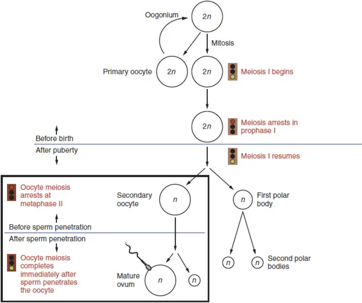
Oogonium: An oogonium is a primordial germ cell found in the fetal ovary, containing a diploid set of chromosomes (2n). These cells undergo mitotic division to increase their numbers before birth, forming the foundation for future egg cells.
Mitosis: Mitosis is a type of cell division where a single cell divides into two identical daughter cells, each with the same number of chromosomes. In oogenesis, oogonia undergo mitosis to produce more oogonia and then differentiate into primary oocytes.
Primary oocyte: A primary oocyte is a diploid cell (2n) that has entered meiosis I but then arrests its development. Females are born with all the primary oocytes they will ever have, each suspended in prophase I.
Meiosis I begins: Meiosis I is the first reductional division of meiosis, where homologous chromosomes separate. This stage marks the beginning of the process that will eventually halve the chromosome number.
Meiosis arrests in prophase I: During fetal development, primary oocytes begin meiosis I but then halt at prophase I. This arrest is maintained until puberty, at which point selected oocytes will resume development each menstrual cycle.
Before birth: This timeframe indicates that the initial stages of oogenesis, including the mitotic proliferation of oogonia and the entry of primary oocytes into meiosis I (followed by arrest), occur during fetal life. A female is born with a finite number of primary oocytes already formed.
After puberty: This signifies the period when oogenesis resumes its progression. From puberty onwards, typically one primary oocyte per menstrual cycle will complete meiosis I and proceed further.
Meiosis I resumes: With the onset of puberty and hormonal stimulation, selected primary oocytes each month resume and complete meiosis I. This division produces two cells of unequal size.
Secondary oocyte: A secondary oocyte is a haploid cell (n) formed after the completion of meiosis I, containing most of the cytoplasm. This cell immediately proceeds into meiosis II but then arrests again.
First polar body: The first polar body is a small, haploid cell (n) that is also formed during the completion of meiosis I, alongside the secondary oocyte. It typically degenerates and does not contribute to fertilization, serving primarily to discard extra chromosomes.
Oocyte meiosis arrests at metaphase II: The secondary oocyte quickly enters meiosis II but then arrests at metaphase II. This second arrest point is crucial and will only be overcome if fertilization occurs.
Before sperm penetration: This denotes the state of the secondary oocyte as it is released from the ovary during ovulation. It remains arrested in metaphase II, awaiting potential fertilization.
After sperm penetration: This crucial event triggers the completion of meiosis II. The entry of a sperm into the secondary oocyte signals the final stages of egg maturation.
Oocyte meiosis completes immediately after sperm penetrates the oocyte: Upon sperm penetration, the secondary oocyte rapidly completes meiosis II. This final division results in the formation of a mature ovum and a second polar body.
Mature ovum: A mature ovum is the final product of oogenesis, a haploid cell (n) containing a complete set of maternal chromosomes. It is now fully prepared for fusion with the sperm nucleus, leading to the formation of a diploid zygote.
Second polar bodies: The second polar body is another small, haploid cell (n) formed during the completion of meiosis II. Like the first polar body, it primarily serves to discard excess genetic material and typically degenerates.
The Remarkable Journey of Oogenesis
Oogenesis is the intricate biological process through which oocytes, or egg cells, are produced in the female body. This journey is remarkably protracted and punctuated by extended periods of arrest, making it a unique example of cell development. Beginning even before birth and culminating only upon fertilization, oogenesis is central to female reproductive capacity and the continuity of species. The diagram provided offers a clear, sequential overview of these stages, highlighting the crucial transitions and checkpoints.
Unlike spermatogenesis, which is a continuous process in males, oogenesis is discontinuous and selective. A female is born with a finite number of primary oocytes, each poised at a specific stage of meiosis. This limited supply underscores the precious nature of female gametes and influences the reproductive lifespan. The careful regulation of this process ensures that only a few oocytes mature each month after puberty, optimizing the chances of successful fertilization.
Key phases and characteristics of oogenesis include:
- Pre-natal proliferation of oogonia and entry into Meiosis I.
- Protracted arrest of primary oocytes in Prophase I until puberty.
- Resumption of Meiosis I and entry into Meiosis II (arrested in Metaphase II) at ovulation.
- Completion of Meiosis II only upon fertilization.
Understanding these stages is not only fundamental to reproductive biology but also provides insight into conditions related to fertility, egg quality, and developmental biology.
From Oogonium to Mature Ovum
The process of oogenesis commences in the fetal ovary with primordial germ cells called oogonia. These diploid cells (2n) undergo numerous mitotic divisions to significantly increase their numbers, ensuring a substantial pool of future egg cells. Subsequently, these oogonia differentiate into primary oocytes, which then embark on meiosis I. However, this meiotic division is swiftly arrested at prophase I, a state that persists until the female reaches puberty. This prolonged arrest means that a female is born with all the primary oocytes she will ever possess, each suspended in this early stage of development.
With the onset of puberty and the cyclic hormonal changes of the menstrual cycle, a select few primary oocytes are stimulated to resume meiosis I each month. Typically, only one will complete this division, resulting in two cells of unequal size: a large secondary oocyte and a small first polar body. The secondary oocyte, now haploid (n) but with duplicated chromosomes, immediately progresses to meiosis II but again arrests, this time at metaphase II. This second arrest is critical, as it signifies the state in which the ovum is released during ovulation. The secondary oocyte will only complete meiosis II if a sperm successfully penetrates its outer layers. Upon fertilization, meiosis II concludes, yielding a mature ovum (haploid, n) and a second polar body. The mature ovum’s nucleus then fuses with the sperm nucleus, forming a diploid zygote, marking the beginning of a new organism. The polar bodies, containing discarded genetic material, typically degenerate.
In conclusion, oogenesis is a meticulously regulated and time-sensitive process that transforms primordial germ cells into a mature ovum capable of fertilization. Its unique characteristic of prolonged arrests ensures the careful management of a finite egg supply over a woman’s reproductive lifetime. This intricate developmental pathway, from fetal development through to fertilization, underscores the remarkable precision of female reproductive biology and its fundamental role in human reproduction.

