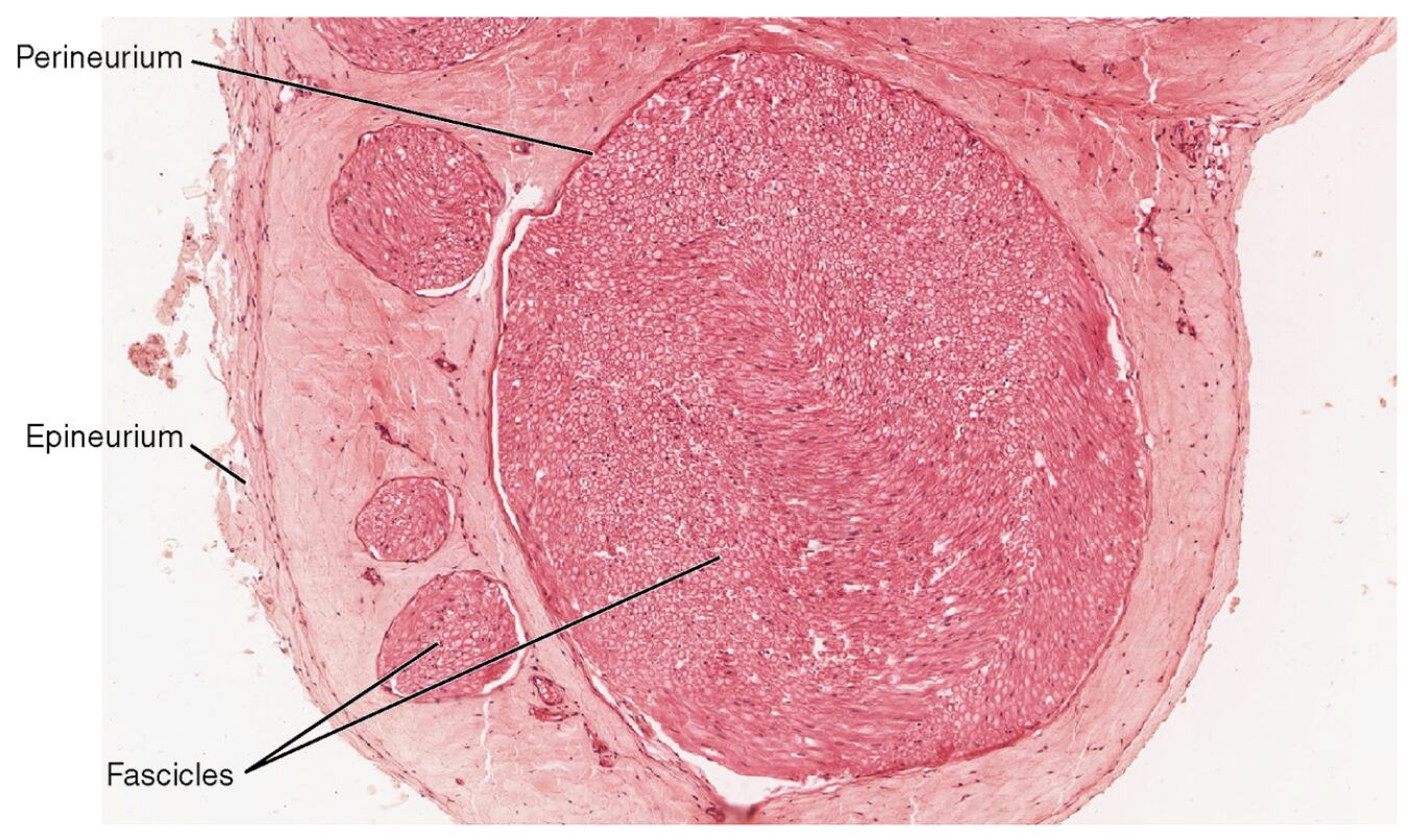
The microscopic view of nerve structure reveals a fascinating organization of connective tissue layers that safeguard and support neural function, as depicted in this detailed image. This exploration into the epineurium, perineurium, and fascicles offers a window into the intricate design that enables nerve impulse transmission and maintains peripheral nerve health. Delving into these components enhances appreciation of the nerve’s resilience and its critical role in the body’s communication network.
Labeled Structures in Nerve Anatomy
This section provides an in-depth look at each labeled component in the microscopic image, explaining their anatomical roles and significance.
Perineurium The perineurium is a multilayered connective tissue sheath encasing each fascicle, providing a protective barrier that maintains a stable internal environment for axons. It contributes to the blood-nerve barrier, preventing harmful substances from reaching the nerve fibers and ensuring optimal function.
Epineurium The epineurium forms the outermost layer of connective tissue surrounding the entire nerve, offering mechanical protection and structural support against external forces. It contains blood vessels and adipose tissue, which supply nutrients and cushion the nerve during movement or injury.
Fascicles The fascicles are bundles of nerve fibers grouped within the perineurium, organized to optimize signal conduction and protect individual axons. These units are essential for grouping fibers with similar functions, such as sensory or motor, enhancing the nerve’s efficiency.
Anatomy of Nerve Structure
The microscopic view highlights the layered organization that defines nerve anatomy. This structure is key to understanding how nerves withstand physical stress while conducting impulses.
- The epineurium, a dense collagenous layer, encases the nerve, providing flexibility and resistance to compression or stretching.
- Fascicles within the nerve are compartmentalized by the perineurium, which also regulates the microenvironment to protect axonal integrity.
- The absence of individual axons in this image focuses attention on the connective tissue framework, though axons are implicitly present within fascicles.
- Blood vessels within the epineurium extend into the perineurium, forming a capillary network that supports the metabolic needs of the nerve.
Physiological Role in Nerve Function
The connective tissue layers play a vital role in supporting nerve impulse transmission and maintaining neural health. Their physiological contributions are essential for effective communication within the nervous system.
- The perineurium’s blood-nerve barrier controls the exchange of ions and molecules, shielding axons from inflammatory or toxic damage.
- The epineurium’s vascular supply ensures a consistent oxygen level, meeting the high metabolic demand of neurons that rely on aerobic respiration.
- Fascicles group fibers with similar functions, such as motor or sensory, allowing for specialized signal processing and transmission.
- This organization supports a conduction velocity that can reach up to 120 m/s in myelinated fibers, depending on axon diameter and myelin thickness.
Clinical Significance and Microscopic Insights
The microscopic perspective offers valuable insights into nerve structure and its clinical relevance. This level of detail aids in diagnosing and managing nerve-related conditions.
- Peripheral nerve injuries affecting the epineurium can lead to neuromas if regeneration is impaired, often necessitating surgical intervention.
- The perineurium’s integrity is crucial in conditions like diabetic neuropathy, where its barrier function may weaken, leading to axonal damage.
- The 40x magnification in this micrograph reveals fascicle arrangement, assisting in the identification of nerve compression or degenerative changes.
- Techniques like nerve conduction studies complement histological findings, helping to assess the functional impact of structural alterations.
Nerve Regeneration and Connective Tissue Support
The connective tissue layers provide a framework for nerve regeneration, a critical process for recovery after injury. This regenerative potential underscores the nerve’s adaptability.
- The epineurium serves as a structural scaffold, guiding regenerating axons with support from its vascular network during the repair phase.
- The perineurium forms bands of Büngner, aligning Schwann cells to direct axonal growth within fascicles after damage.
- Successful regeneration depends on minimizing scar tissue within the epineurium, which can obstruct axonal regrowth if excessive.
- Growth factors released by surrounding cells enhance this process, promoting the restoration of nerve function over time.
In conclusion, the microscopic view of nerve structure illuminates the sophisticated interplay of connective tissue layers that protect and sustain neural activity. The detailed anatomy of the epineurium, perineurium, and fascicles provides a foundation for advancing our understanding of nerve function, offering insights that support improved diagnostic and therapeutic approaches in clinical practice.

