The muscular artery, a vital link in the circulatory system, delivers oxygenated blood to specific organs and tissues, adapting to varying metabolic demands with its robust design. This image highlights the tunica intima, tunica media, tunica adventitia, and smooth muscle cells, showcasing the structural features that enable these medium-sized vessels, such as the brachial or femoral arteries, to regulate blood flow effectively.
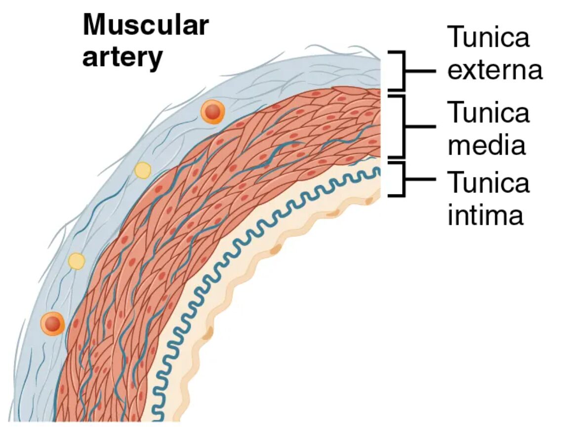
Tunica intima Tunica intima is the innermost layer, consisting of a single layer of endothelial cells that line the lumen and minimize friction during blood flow. This layer includes a subendothelial layer and an internal elastic lamina, which provides some elasticity to accommodate pressure changes.
Tunica media Tunica media forms the thick middle layer, dominated by circular smooth muscle cells with fewer elastic fibers compared to elastic arteries. This structure allows precise control of vessel diameter, enabling the artery to adjust blood flow to meet the metabolic needs of specific organs.
Tunica adventitia Tunica adventitia is the outer layer, composed of connective tissue and collagen fibers that anchor the artery to surrounding tissues. This layer is thinner than in elastic arteries but provides essential support and contains vasa vasorum to nourish the vessel wall.
Smooth muscle cells Smooth muscle cells are the primary component of the tunica media, appearing as elongated cells under magnification, and are responsible for vasoconstriction and vasodilation. These cells respond to neural and hormonal signals, fine-tuning blood distribution to active tissues.
The Role of Muscular Artery Layers in Circulation
This analysis reveals how each layer supports the muscular artery’s regulatory function. The design ensures efficient blood delivery to specific regions of the body.
- Tunica Intima Function: The endothelial cells release nitric oxide to promote vasodilation, preventing clot formation. The internal elastic lamina offers flexibility, accommodating pulsatile flow.
- Tunica Media Role: The smooth muscle cells contract or relax to adjust lumen size, controlled by the sympathetic nervous system. This adaptability directs blood to organs like the kidneys or muscles.
- Tunica Adventitia Support: The collagen network stabilizes the artery, preventing overexpansion under pressure. The vasa vasorum supplies nutrients to the thicker media layer.
- Smooth Muscle Cell Activity: These cells respond to adrenaline, increasing vasoconstriction during stress. Their coordinated action maintains optimal blood pressure.
Anatomical Details of Muscular Arteries
The image provides a detailed view of the muscular artery’s layered structure, emphasizing its muscular dominance. The thickness of the tunica media stands out in this design.
This close-up perspective aids in understanding the artery’s adaptability. It serves as a foundation for studying vascular anatomy and physiology.
- Tunica Intima Composition: The endothelium is supported by a thin subendothelial layer, with the internal elastic lamina less prominent than in elastic arteries. This layer varies slightly with vessel size.
- Tunica Media Thickness: The layer contains multiple layers of smooth muscle cells, with fewer elastic lamellae than elastic arteries. This muscular focus is ideal for active diameter control.
- Tunica Adventitia Variation: Collagen fibers form a supportive mesh, with the vasa vasorum visible as small vessels. This layer is thinner, reflecting the artery’s medium size.
- Smooth Muscle Cell Structure: These cells appear as spindle-shaped units, arranged circularly around the lumen. Their density increases toward the outer media layer.
Physiological Functions of Muscular Artery Layers
The physiological roles of these layers are tailored to regulate blood flow to specific tissues. Their design ensures effective oxygen and nutrient delivery.
Each layer contributes uniquely to arterial performance. This functionality supports the body’s cardiovascular demands.
- Blood Flow Regulation: The tunica media’s smooth muscle adjusts lumen size, responding to metabolic signals like low oxygen. This control directs blood to active tissues during exercise.
- Pressure Maintenance: The internal elastic lamina in the tunica intima smooths the pulse wave, maintaining pressure around 100-120 mmHg. This protects downstream vessels.
- Oxygen Delivery: The artery carries oxygenated blood rich in nutrients like glucose, facilitated by the tunica intima’s smooth surface. This ensures efficient supply to organs.
- Vasomotor Control: Smooth muscle cells in the tunica media respond to acetylcholine, promoting vasodilation. This fine-tunes blood distribution based on tissue needs.
Comparative Anatomy with Other Arterial Types
The muscular artery contrasts with elastic arteries and arterioles, reflecting its unique role. This image emphasizes these structural differences.
The visual representation highlights the muscular artery’s distinct features. This understanding is key to grasping vascular diversity.
- Muscular vs. Elastic Arteries: Muscular arteries have more smooth muscle and fewer elastic lamellae, unlike the elastic-dominated media of elastic arteries. This reflects their role in distribution versus pressure maintenance.
- Muscular vs. Arterioles: Muscular arteries feature a thicker tunica media, while arterioles have a thinner, more contractile layer. This shift supports finer flow control.
- Transition to Arterioles: The tunica media thins as muscular arteries branch into arterioles, reducing muscle layers. This gradient adapts to changing pressure and resistance.
- Histological Features: Staining reveals dense smooth muscle bands in the tunica media, contrasting with the elastic focus in elastic arteries. This aids in microscopic identification.
Clinical and Research Perspectives
Insights from muscular artery anatomy inform medical practice and research. The layered structure is a focus for studying cardiovascular health and disease.
Advances in imaging and histology enhance these studies, offering diagnostic tools. These efforts support innovative treatment strategies.
- Atherosclerosis Risk: Plaque in the tunica intima narrows the lumen, common in muscular arteries like the coronary artery. Microscopic analysis guides statin therapy.
- Hypertension Impact: Thickened tunica media in response to chronic pressure reduces elasticity. This finding informs antihypertensive treatments.
- Peripheral Artery Disease: Reduced blood flow due to smooth muscle dysfunction affects muscular arteries. Imaging detects early narrowing for intervention.
- Therapeutic Innovations: Targeting smooth muscle with calcium channel blockers treats hypertension. Stem cell research explores regenerating arterial layers.
In conclusion, this image of a muscular artery provides a detailed look at the tunica intima, tunica media, tunica adventitia, and smooth muscle cells, revealing their critical roles in regulating blood flow. These anatomical insights not only deepen our understanding of arterial function but also support advancements in diagnosing and treating cardiovascular conditions.

