The muscles of facial expression are a remarkable feature of human anatomy, enabling the diverse range of emotions we display through movements of the face. This lateral view image showcases key muscles that insert into the skin around the eyelids, nose, and mouth, facilitating expressions by moving the skin rather than bones. Delving into this anatomy offers a deeper understanding of how these muscles contribute to both communication and facial aesthetics.
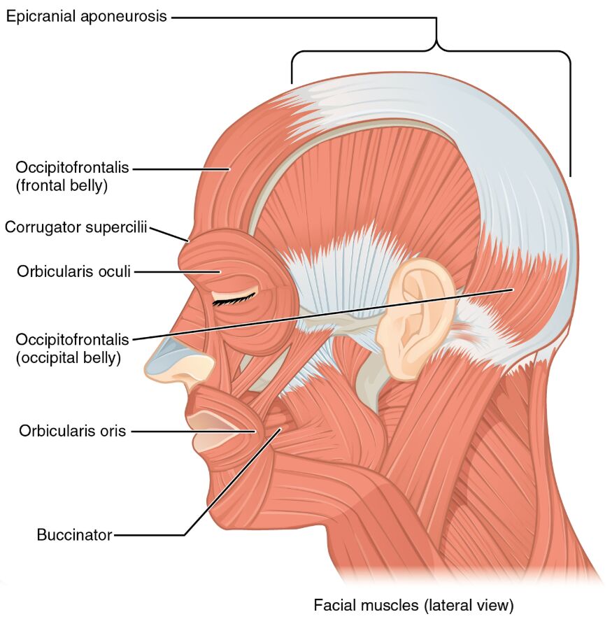
Epicranial aponeurosis
The epicranial aponeurosis is a dense, fibrous layer that spans the top of the skull, serving as an attachment point for the occipitofrontalis muscle. It plays a vital role in transmitting muscle forces across the scalp, ensuring smooth and coordinated movements.
Occipitofrontalis (frontal belly)
The occipitofrontalis, with its frontal belly located on the forehead, raises the eyebrows and creates forehead wrinkles, often linked to expressions of surprise or curiosity. This muscle connects to the epicranial aponeurosis, enhancing its ability to move the scalp.
Corrugator supercilii
The corrugator supercilii lies beneath the medial eyebrow and draws the eyebrows downward and inward, forming vertical wrinkles above the nose. This action is commonly associated with frowning or concentration, reflecting its role in emotional expression.
Orbicularis oculi
The orbicularis oculi encircles the eye, enabling the closure of the eyelids for blinking or tight squinting during strong emotions. Its sphincter-like function protects the eye and supports expressions like winking or crying.
Occipitofrontalis (occipital belly)
The occipital belly of the occipitofrontalis, located at the back of the head, pulls the scalp backward, working in opposition to the frontal belly. This movement can subtly influence the tension across the scalp and contribute to certain facial expressions.
Orbicularis oris
The orbicularis oris surrounds the mouth, acting as a sphincter to close the lips and facilitate actions like kissing or speaking. Its intricate structure allows for precise control, making it essential for a wide range of lip movements.
Buccinator
The buccinator, situated in the cheek, compresses the cheeks to assist in chewing by pushing food between the teeth. It also supports actions like blowing air or sucking, contributing to both function and facial contour.
The muscles of facial expression, as depicted in this lateral view, are a cornerstone of human anatomy, uniquely designed to interact with the skin to produce a vast array of expressions. This image provides a clear perspective on their arrangement and function, serving as an invaluable resource for exploring their role in daily life. By examining their structure and interplay, one can better appreciate the complexity behind every facial movement and its significance in non-verbal communication.
Anatomical Overview of Facial Expression Muscles
Facial expression muscles offer a fascinating study due to their distinctive structure and function. These muscles, innervated by the facial nerve (cranial nerve VII), originate from bony structures or fascia and insert into the skin, allowing for dynamic facial movements.
- Diverse origins: The occipitofrontalis (frontal belly) arises from the epicranial aponeurosis, while the occipital belly connects to the occipital bone.
- Skin insertion: Their attachment to the dermis enables direct skin movement, a key difference from muscles that move bones.
- Nerve supply: The facial nerve provides precise control, coordinating actions like smiling or frowning.
- Vascular support: The facial artery and its branches ensure a steady blood supply, supporting the muscles’ constant activity.
This lateral view highlights the spatial relationships between these muscles, offering insight into their collaborative efforts. Their unique anatomy underpins the versatility of facial expressions, making them a critical area of study.
Functions and Movements in the Lateral Perspective
Each muscle in the lateral view plays a specific role in facial expression. Their coordinated actions create the emotional landscape we recognize on the human face.
- Epicranial aponeurosis function: Acts as a tendon, distributing forces from the occipitofrontalis to stabilize scalp movements.
- Occipitofrontalis (frontal belly) action: Elevates the eyebrows, producing forehead wrinkles associated with surprise.
- Corrugator supercilii movement: Draws the eyebrows together, forming frown lines that indicate concentration or displeasure.
- Orbicularis oculi role: Closes the eyelids, protecting the eye and enabling expressions like squinting.
- Occipitofrontalis (occipital belly) contribution: Pulls the scalp backward, balancing the frontal belly’s action.
- Orbicularis oris function: Closes and puckers the lips, essential for speech and emotional displays.
- Buccinator activity: Compresses the cheeks, aiding chewing and supporting actions like blowing.
These movements showcase the muscles’ adaptability, responding to both voluntary commands and involuntary reflexes. Their interplay ensures a seamless range of expressions critical for human interaction.
Clinical Significance of Facial Muscles
Knowledge of these muscles has practical applications in clinical practice. Dysfunction or injury can lead to significant functional and aesthetic challenges, requiring expert intervention.
- Facial nerve injury: Damage can paralyze muscles like the orbicularis oculi, impairing eyelid closure.
- Bell’s palsy impact: This condition affects muscles like the buccinator, causing unilateral facial weakness.
- Surgical considerations: Reconstructive procedures rely on understanding muscle placement for symmetry restoration.
- Cosmetic use: Botox targets muscles like the corrugator supercilii to reduce frown lines, demanding anatomical precision.
These examples emphasize the importance of mastering facial muscle anatomy for effective treatment. Accurate diagnosis and intervention can greatly enhance patient recovery and quality of life.
Conclusion
The muscles of facial expression, as illustrated in this lateral view, reveal the intricate design behind human emotional expression. Their ability to move the skin rather than bones allows for the rich variety of facial movements that define our interactions. This guide provides a thorough exploration of their anatomy, function, and clinical relevance, offering a solid foundation for further study. By understanding the roles of the epicranial aponeurosis, occipitofrontalis, and others, one can gain a deeper appreciation for the mechanisms driving every smile or frown.

