Veins are crucial vessels in the circulatory system, responsible for returning deoxygenated blood to the heart, and their microscopic structure reveals the intricate layers that support this function. This image provides a histological section of a vein, showcasing its anatomical features as observed under a microscope, offering a window into the cellular organization that ensures efficient blood flow.
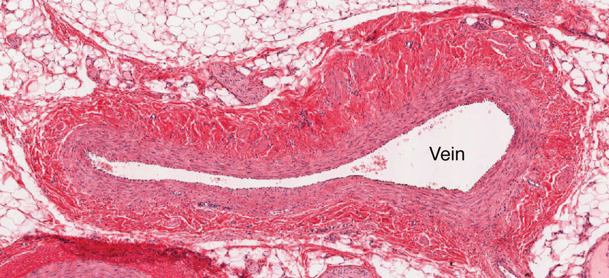
Vein The vein is the primary structure depicted, identifiable by its large, thin-walled lumen surrounded by multiple tissue layers. This vessel facilitates the low-pressure return of blood to the heart, adapting to varying volumes through its flexible walls.
The Role of Veins in the Circulatory System
Veins play an essential role in maintaining blood circulation and volume regulation. Their structure supports the body’s ability to adapt to physiological changes.
- The vein acts as a reservoir, holding a significant portion of the body’s blood supply.
- Its thin walls allow it to expand and contract, accommodating changes in blood volume.
- Veins work in conjunction with the skeletal muscle pump to aid venous return.
- They transport deoxygenated blood, including hormones like T3 and T4 from the thyroid gland, back to the heart.
Anatomical Features of Veins Under the Microscope
The microscopic view of a vein reveals a complex arrangement of tissue layers. These layers contribute to the vein’s functionality and resilience.
- The vein lumen appears large and irregular, reflecting its role in low-pressure circulation.
- Surrounding tissues include layers of smooth muscle and connective tissue for support.
- Endothelial cells line the interior, reducing friction during blood flow.
- The overall structure contrasts with arteries due to its thinner muscular layer.
Physiological Functions and Importance
The microscopic anatomy of veins enables them to perform critical physiological roles. This structure ensures efficient blood transport and adaptation to bodily demands.
- The vein supports the return of blood against gravity, especially in the lower limbs.
- Its flexibility aids in maintaining venous pressure during changes in posture.
- The vessel facilitates the removal of metabolic waste from tissues.
- This adaptability is vital for overall cardiovascular health.
Clinical Significance of Vein Microstructure
Understanding the microscopic structure of veins provides insights into potential health issues. Alterations in these tissues can indicate or contribute to circulatory disorders.
- Thickening of the vein wall may signal chronic venous insufficiency, affecting blood flow.
- Damage to the endothelial lining can increase the risk of thrombosis.
- Varicose veins may develop due to weakened structural support in the vein.
- Research into vein histology supports advancements in vascular treatments.
Comparison with Other Blood Vessels
Veins differ from arteries and capillaries due to their specific microscopic features. This comparison highlights their unique role in the circulatory system.
- Unlike arteries, the vein has a thinner tunica media, reflecting lower pressure demands.
- Capillaries lack the layered structure seen in veins, focusing on exchange rather than transport.
- Veins contain valves in some regions, absent in this section but critical for function.
- The vein‘s larger lumen contrasts with the narrower capillaries.
Maintenance and Regulation of Veins
The body employs mechanisms to maintain the health and function of veins. These processes ensure optimal performance under varying conditions.
- Endothelial cells in the vein release substances to regulate blood flow and prevent clotting.
- Connective tissue provides ongoing support, adapting to mechanical stress.
- Neural and hormonal signals influence vein diameter and tone.
- This regulation supports efficient venous return and tissue perfusion.
In conclusion, the microscopic structure of a vein, as shown in this image, illustrates the remarkable design that enables efficient blood return to the heart. With its large lumen and supportive tissue layers, this vessel exemplifies the circulatory system’s ability to adapt to diverse physiological needs. Exploring these features enhances our understanding of venous anatomy and its critical role in maintaining overall health.

